
Researched by Consultants from Top-Tier Management Companies

Powerpoint Templates
Icon Bundle
Kpi Dashboard
Professional
Business Plans
Swot Analysis
Gantt Chart
Business Proposal
Marketing Plan
Project Management
Business Case
Business Model
Cyber Security
Business PPT
Digital Marketing
Digital Transformation
Human Resources
Product Management
Artificial Intelligence
Company Profile
Acknowledgement PPT
PPT Presentation
Reports Brochures
One Page Pitch
Interview PPT
All Categories

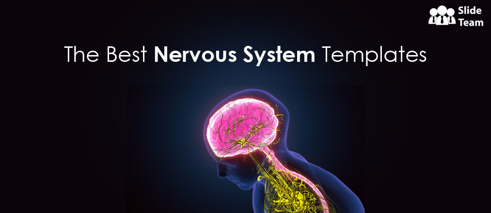
The Nervous System in 17 Templates
Lakshya Khurana
Take a moment to think about this: You are the brain. Your pains and pleasures, happiness and sadness, dreams and aspirations, fears and doubts, love and hate, every feeling and emotion, everything that makes you YOU, are within some three pounds of gray matter in your head.
Take a moment to think about it.
It’s not exactly a brain-in-a-jar, but it’s not that far off either (and how would we know otherwise?). The brain doesn’t do it alone, of course. It has the spinal cord, the nerves, some chemistry, and a jolt of electricity to help. This is what this blog is going to talk about, the NERVOUS SYSTEM, which lets you experience the world around you.
This blog offers 17 gorgeous PowerPoint Templates to give you the best presentation on this human organ system.
Whether you are a high school student, a medical student, or a doctor, it has no bearing on whether this collection is right for you because the templates are:
- CONTENT-READY: You do not need to conduct any research; we have curated that for you. Should you be running late, your presentation is there for you.
- EDITABLE: Any information that catches your attention can be added to or deleted from these slides.
Each deck has several slides presenting the diagram with and without the labels and highlighting individual parts, making for a world-class comprehensive presentation!
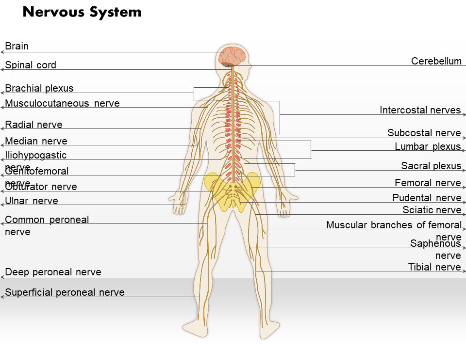
Template 1: The Nervous System
This PPT Deck covers the structure and function of the nervous system and its role in human health and disease. It provides detailed, easy-to-understand information and is an excellent resource for students and professionals alike. Download it now by clicking the link below.
Download this template
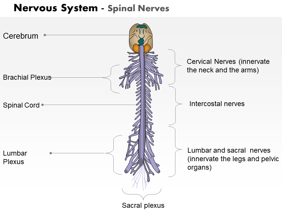
Template 2: The Nervous System - Spinal Nerves
This PowerPoint Set is an essential tool for anyone interested in learning more about the human nervous system. This PowerPoint Presentation covers everything from the anatomy of the spinal cord to the functions of neurons. Get it right away!
Grab this template
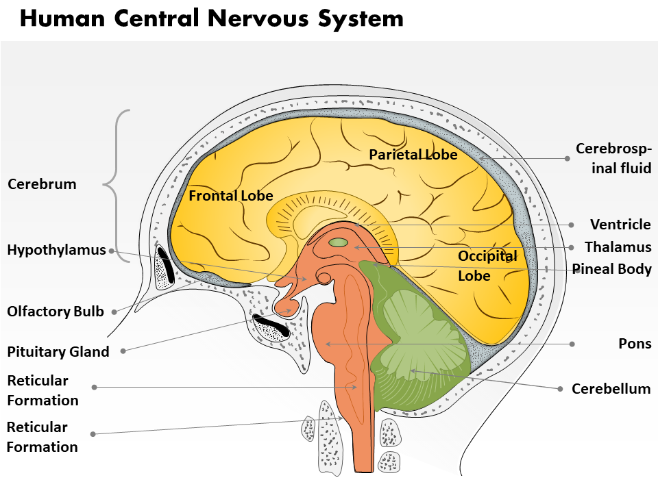
Template 3: How Relationships Work in the Nervous System
With this PPT Bundle, you can showcase topics related to the cerebrum, hypothalamus, parietal lobe, pituitary gland, reticular formation, cerebellum, etc, in your presentation. Plus, the clear and concise layout will help your audience understand the relationships between different brain parts. Make it yours now.
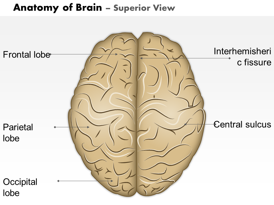
Template 4: Anatomy of the Brain - Superior View and Piecing the Jigsaw Together
This detailed PowerPoint Layout provides a comprehensive overview of the brain and its functions. Every major area of the brain is labeled and explained. from the frontal lobe to the central sulcus. The superior view allows you to see the parts of the brain and how they fit together. Download it now.
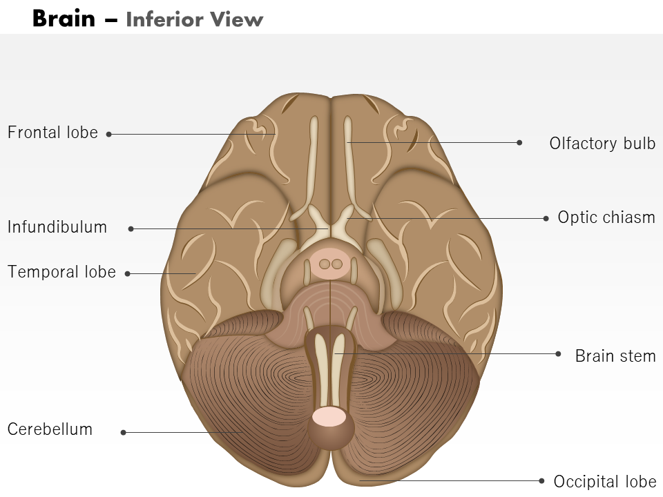
Template 5: Anatomy of the Brain – Peak into intricacies of existence with Inferior View
This PPT Preset is a comprehensive resource that provides complete information about the structure and function of the brain. It covers the brain’s anatomy from an inferior view, allowing students to visualize this important organ’s intricacies. Get it right away!
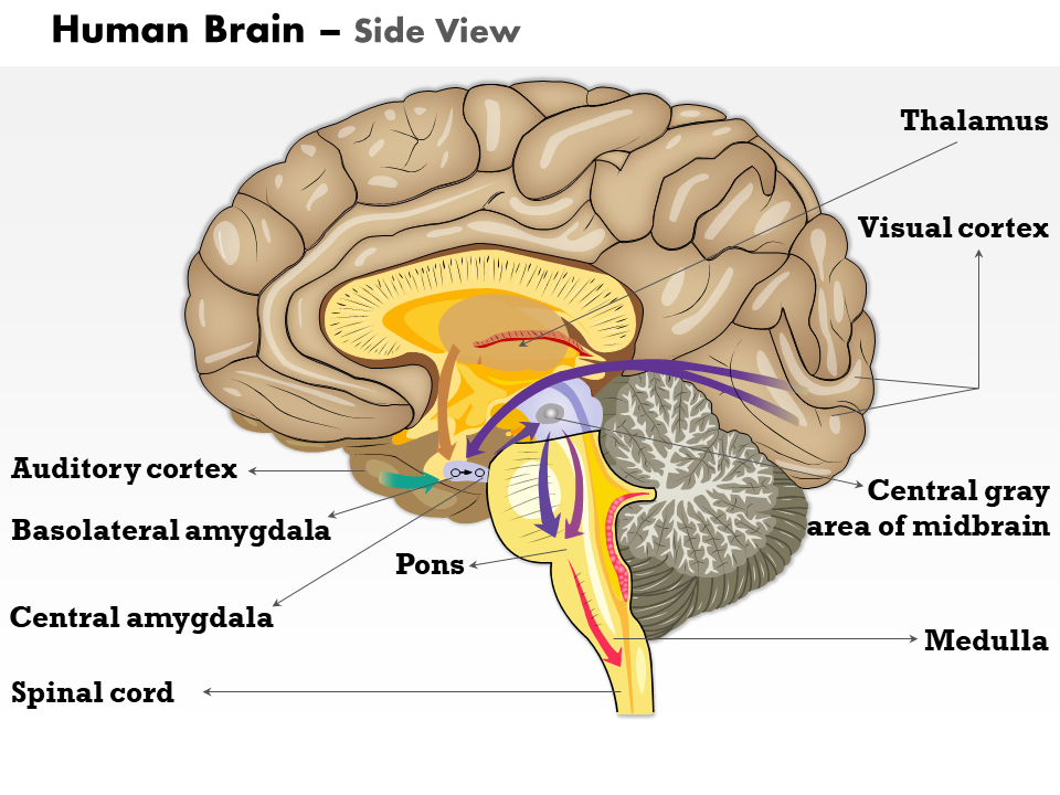
Template 6: Anatomy of the Brain – Peak into intricacies of existence with Side View
This PowerPoint Deck is packed with information on parts of the brain, such as the pons, medulla, visual cortex, etc, as well as how they function together. And to top it off, it's also visually stunning, with beautiful illustrations that bring the subject matter to life. Make it yours, now!
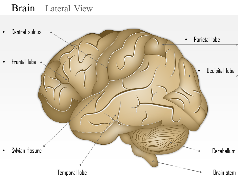
Template 7: Anatomy of the Brain – Get the corner seat with Lateral View
Use this PPT Set to showcase the brain’s lateral view and highlight the parts like cerebellum, Sylvian fissure, temporal lobe, and more. Yet another important view of the brain, download it now by clicking the link below.
Template 8: Anatomy of the Brain – Split and Deliver in Sagittal View
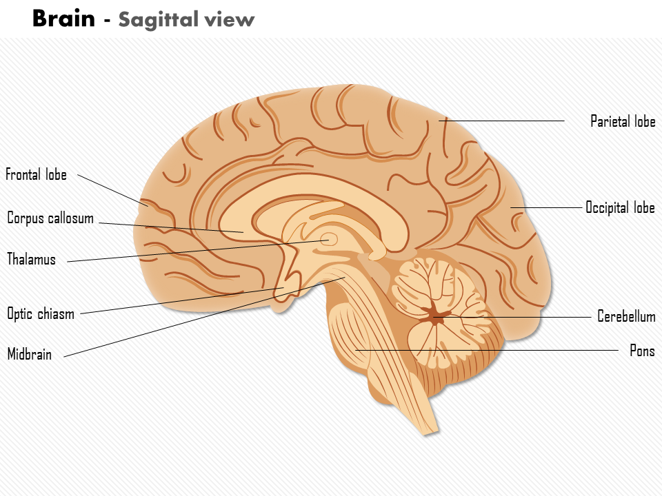
A unique look into the brain’s anatomy, the sagittal view splits the organs left and right. Present the hidden parts of the brain, such as the optic chiasm and thalamus, among others. Get this PowerPoint Bundle now.
Template 9: Anatomy of the Brain – The Hard Reality of Skull and Coronal View
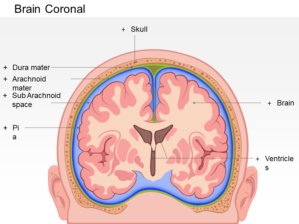
Showcase and explain how the brain resides inside the skull and the layers that cover it with this PPT Layout. The brain has its own system of interacting with other organs, such as the blood-brain barrier and the spinal fluid. Get this deck now.
Template 10: The Cerebellum: Secret to Maintaining Body Balance
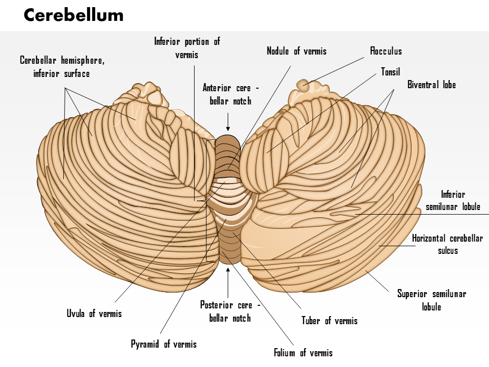
The cerebellum is a small, round structure located at the back of the brain. It is responsible for coordinating movement and balance. The cerebellum is divided into two hemispheres, each of which controls the opposite side of the body. All this information and more is within this PowerPoint Preset. Download now.
Template 11: The Spinal Cord: The VIP Needs Protection
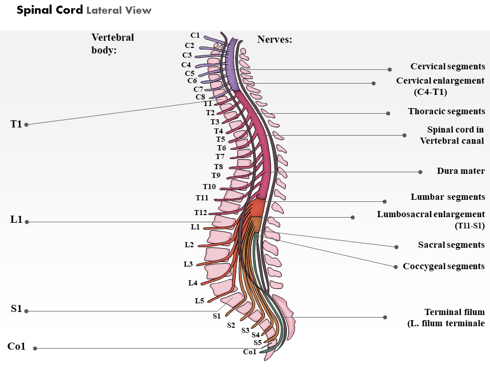
This PPT Deck presents the spinal cord, which is a vital part of the central nervous system. It extends from the brainstem down to the lower back, with the vertebral column protecting it. Detail the parts of this important organ and educate the audience on keeping it safe. Download it now.
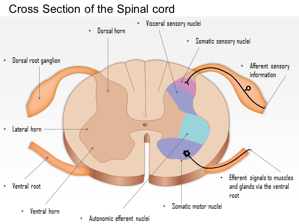
Template 12: Cross-Section of the Spinal Cord: Movement and health
A diagram of the spinal cord can be an invaluable tool for understanding this complex structure and how it works. It shows parts of the spinal cord and how they are arranged. It is an essential tool for students of anatomy, physiotherapy, or any other discipline concerned with human health. Make this PowerPoint Set yours with a download.
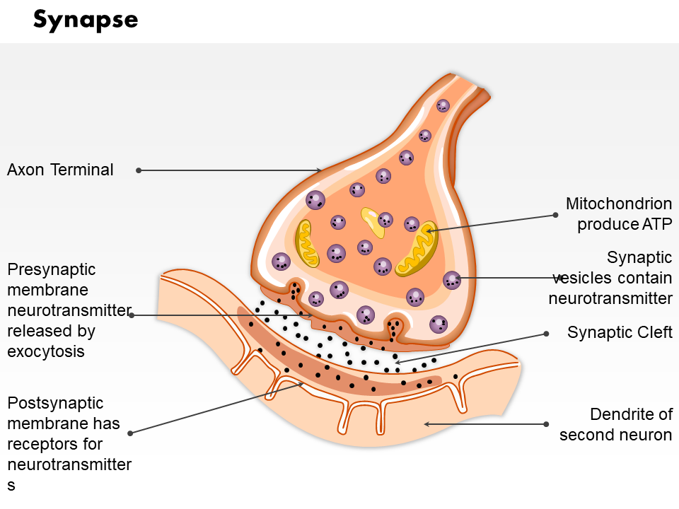
Template 13: The Synapse: The Medium that Lets You Say
Are you looking for a way to better understand how the brain works? A synapse diagram can be a helpful tool in doing just that. Synapses are the communication points between neurons, and this PPT Bundle provides a clear and concise visual representation of how they work. Get it now.
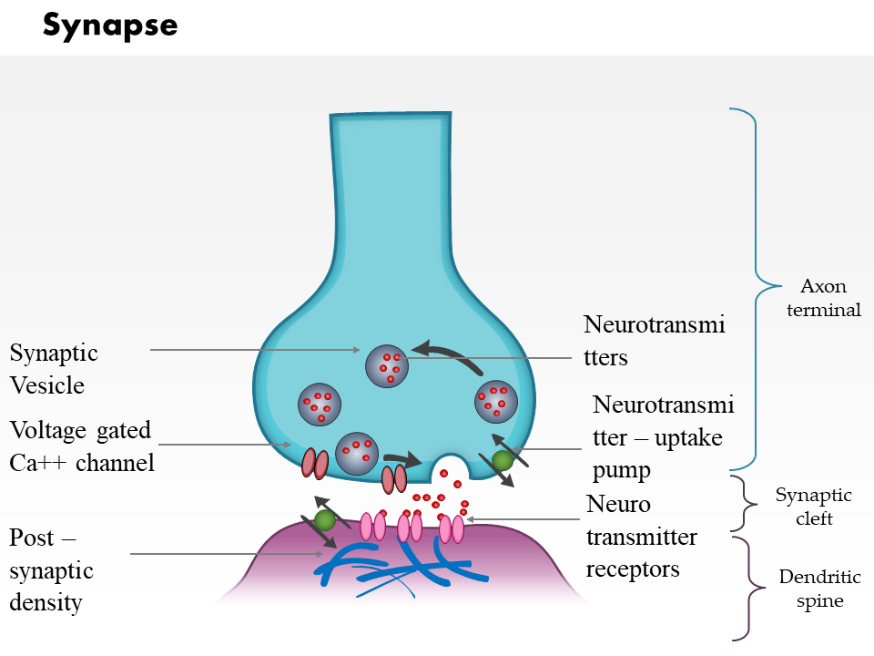
Template 14: Synapse 2 - Chemistry Involved in Transmission
The synapse chemistry diagram is the perfect tool for studying and understanding the chemical processes that take place at a synapse. This PowerPoint Preset shows the structures and functions of molecules involved in synaptic transmission. Download it now.
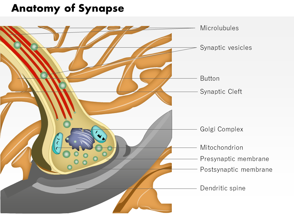
Template 15: Synapse 3: The Anatomy: The Powerhouse in Action
This PPT layout presents a detailed diagram of the synaptic cell, with its organelles like the mitochondria (the powerhouse of the cell), Golgi complex, microlubules, etc. Download this template now by clicking the link below.
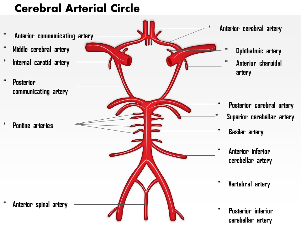
Template 16: The Cerebral Arterial Circle: Key to Genuine Understanding
The Cerebral Arterial Circle diagram is a highly detailed and accurate representation of the arteries that supply blood to the brain. It is essential to understand if you wish to understand the brain. This PowerPoint deck presents the artery diagram with labels. Get it now.
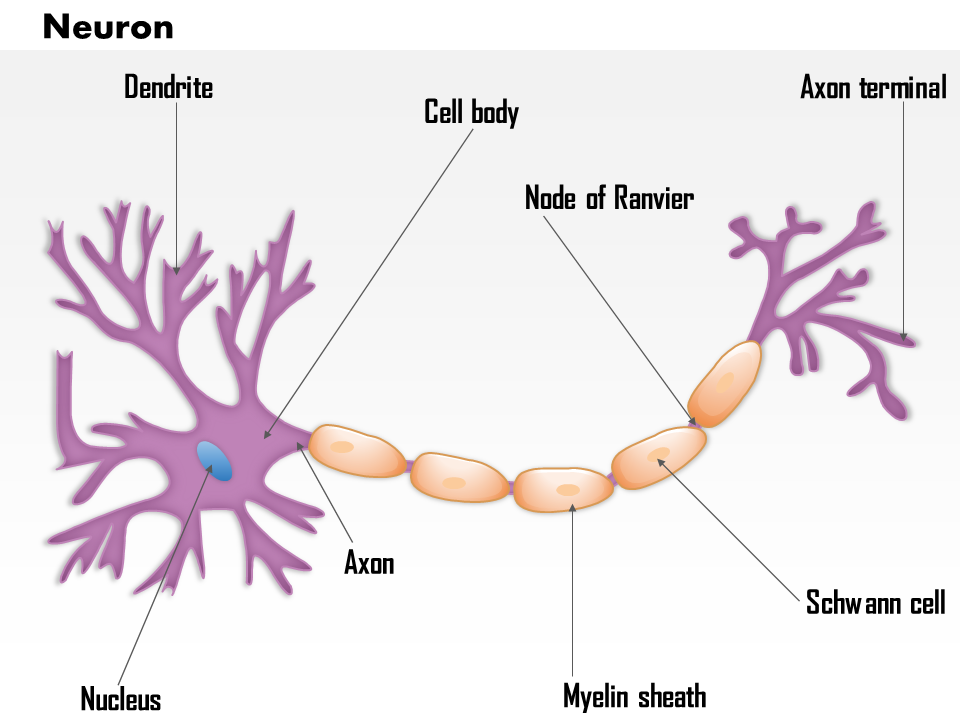
Template 17: The Neuron: Signals that Matter
The neuron is a specialized cell of the nervous system that is responsible for transmitting signals throughout the body. Use this PPT set to label and explain the structure of the cell and how it functions. Download it now.
Complexity simplified
The nervous system is an incredibly complex system, and scientists are still learning a great deal about how it works. However, we know it plays a vital role in maintaining our health and well-being. Without it, we would not be able to interact with our environment or make sense of the world around us.
You can also check out our blog with eight amazing templates on the cardiovascular system !
A Refresher
The following text is written to provide you with a refresher on the nervous system, its function, and how it works. We'll cover everything from the brain and its lobes to the nerves and how they transmit signals throughout the body.
FAQs for the Nervous System
How does the nervous system work.
The nervous system is responsible for sending signals throughout the body and consists of the brain, spinal cord, and nerves. The brain is the control center for the nervous system, and the spinal cord carries messages between the brain and the rest of the body. Nerves are responsible for carrying signals to and from the brain and spinal cord.
What is the nervous system made up of?
The nervous system comprises two parts: the central nervous system (CNS) and the peripheral nervous system (PNS). The CNS consists of the brain and the spinal cord. The PNS consists of all the nerves that branch off from the brain and spinal cord.
What does the nervous system do?
The nervous system is responsible for the regulation and coordination of body functions. It transmits signals between parts of the body and controls the activities of all other organs. It is like a command center of the human body, where, at any moment, a lot of frenetic activity is on.
What is the peripheral nervous system?
The peripheral nervous system is the part of the nervous system comprising all nerves and ganglia outside of the brain and spinal cord. Its main function is to connect the central nervous system to the rest of the body.
The peripheral nervous system can be divided into two main parts: the somatic nervous system and the autonomic nervous system. The somatic nervous system is responsible for carrying information from the body to the brain, and the autonomic nervous system is responsible for carrying information from the brain to the body.
What is the autonomic nervous system?
The autonomic nervous system controls involuntary body functions, such as your heart rate and blood pressure. It's made up of two divisions, the sympathetic nervous system and the parasympathetic nervous system. Both systems work together to ensure your body functions properly.
What does the sympathetic nervous system do?
The sympathetic nervous system is responsible for the "fight-or-flight" response. This occurs when the body perceives a threat and releases adrenaline and other hormones to prepare the body for action. The sympathetic nervous system speeds up the heart rate, increases blood pressure, and diverts blood flow to the muscles. It also increases blood sugar levels and dilates the pupils of the eyes.
What does the parasympathetic nervous system do?
The parasympathetic nervous system is responsible for the "rest and digest" response in the body. This means that the parasympathetic nervous system is active when the body is at rest. This system slows down the heart rate and increases the activity of the digestive system. The parasympathetic nervous system is also responsible for maintaining the body's blood pressure and blood sugar levels.
Related posts:
- How to Design the Perfect Service Launch Presentation [Custom Launch Deck Included]
- Quarterly Business Review Presentation: All the Essential Slides You Need in Your Deck
- [Updated 2023] How to Design The Perfect Product Launch Presentation [Best Templates Included]
- 99% of the Pitches Fail! Find Out What Makes Any Startup a Success
Liked this blog? Please recommend us

Top 15 Healthcare Brochure Templates to Advertize Your Medical Services

Top 20 PowerPoint Templates to Create a Fast-Acting Clinical Development Plan
This form is protected by reCAPTCHA - the Google Privacy Policy and Terms of Service apply.

Digital revolution powerpoint presentation slides

Sales funnel results presentation layouts
3d men joinning circular jigsaw puzzles ppt graphics icons

Business Strategic Planning Template For Organizations Powerpoint Presentation Slides

Future plan powerpoint template slide

Project Management Team Powerpoint Presentation Slides

Brand marketing powerpoint presentation slides

Launching a new service powerpoint presentation with slides go to market


Agenda powerpoint slide show

Four key metrics donut chart with percentage

Engineering and technology ppt inspiration example introduction continuous process improvement

Meet our team representing in circular format

Home PowerPoint Templates Template Backgrounds Nervous System PowerPoint Template
Nervous System PowerPoint Template
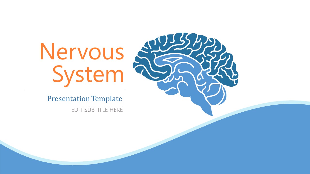
The Nervous System PowerPoint Template presents illustrative diagrams for neuroscience theme concepts. It provides engaging graphics and descriptive labels for medical analysis and academic presentations. You can create a highly informative presentation using the nervous system clean PowerPoint templates. For educational and medical purposes, the medical PowerPoint template diagrams demonstrate the structure of a nervous system. Use pre-design professional PowerPoint templates to boost your presentation session about neurology.
Pharmaceuticals or medical equipment manufacturers can use slide presentation templates to assess research projects with scientific studies. PPT presentation templates are ideal PowerPoint to discuss the anatomy of the human nervous system. The use of labels helps describe the physical location of the body or cell parts. Academic professionals can benefit from these templates to teach biology and medical education. The professional presentation PowerPoint templates of neurons are animated diagrams to explain the functions and components of nerve cells.
The nervous system is the most important biological system in the human body to transmit sensory information. The nervous system has two parts: the central nervous system and the peripheral nervous system. The healthcare PowerPoint templates provide diagrams for both CNS and PNS along with brain and neuron diagrams. A neuron is a nerve cell that transmits information to other nerve cells.
The Nervous System PowerPoint Template contains drawings of the brain, neuron, and nervous system. Editable PowerPoint templates let users customize the contents of each nervous system diagram with relevant descriptions. PowerPoint shapes can be copied to other slide designs or Google Slides Template for ease of presentation. Simple PowerPoint backgrounds make it easier to copy slides into any animated PowerPoint themes. Users can also change professional PowerPoint backgrounds color or change the arrangement of nervous system graphics.
You must be logged in to download this file.
Favorite Add to Collection
Details (9 slides)

Supported Versions:
Subscribe today and get immediate access to download our PowerPoint templates.
Related PowerPoint Templates

Animated Student Intro PowerPoint Template

Incident Report PowerPoint Template

Workshop Template PowerPoint

Book Report Presentation Template
ANATOMY OF THE NERVOUS SYSTEM
Jan 03, 2020
680 likes | 901 Views
ANATOMY OF THE NERVOUS SYSTEM. Lecture 9 &10. Functions of the nervous system. 1. Initiate and/or regulate movement of body parts 2. Regulate secretions from glands 3. Gather information about the external environment and the internal environment of the body
Share Presentation
- nervous system
- intermediate zone
- neural tube
- ventral intermediate zone
- intermediate zone comprises neurons

Presentation Transcript
ANATOMY OF THE NERVOUS SYSTEM Lecture 9 &10
Functions of the nervous system • 1. Initiate and/or regulate movement of body parts • 2. Regulate secretions from glands • 3. Gather information about the external environment and the internal environment of the body • using senses (sight, hearing, touch, balance, taste) and mechanisms to detect pain, temperature, pressure, and certain chemicals, such as carbon dioxide, hydrogen, and oxygen • 4. Maintain an appropriate state of consciousness • 5. Stimulate thirst, hunger, fear, rage, and sexual behaviors appropriate for survival
The entire nervous system can be divided into two parts: • 1- Central nervous system (CNS) • includes the brain and spinal cord • 2- Peripheral nervous system (PNS), which consists of: • Cranial nerves • spinal nerves
A further distinction is often made: • the autonomic nervous system (ANS) • which integrates activity of visceral structures • smooth muscle, cardiac muscle, and glands • The ANS has elements in both: • Central nervous system. • Peripheral nervous system.
In the PNS: 1- sensory (afferent) nerves - gather information about the external and internal environments and relay this information to the CNS. - The specialized organs or cells that detect specific stimuli are sensory receptors.
CNS interprets information arriving via the PNS, integrates that information, and initiates • appropriate movement of body parts • glandular secretion • behavior response. 2- Motor efferent - Communication between the CNS and muscles and accomplished via nerves of the PNS
Microscopic Neuroanatomy • The individual nerve cell is called a neuron. • Each neuronal cell body gives rise to one or more nerve processes and cytoplasmic extensions of the cell. • The nerve processes are called dendritesif they transmit electrical signals toward the cell bodies • They are called axonsif they conduct electrical signals away from the cell bodies.
Neurons may be classified morphologically according to their number of nerve processes: • Unipolar neuronshave one process • Bipolar neuronshave one dendrite and one axon • Multipolar neuronshave a number of dendrites in addition to their single axon. • We do not have true unipolar neurons, but many sensory neurons have their single dendrite and axon fused so as to give the appearance of a single process • This configuration is pseudounipolar.
Nervous tissue consists not only of neurons but also of supportive cells. • In the CNS, these supportive cells are the neuroglia, comprising a variety of glial cells • Groups of nerve cell bodies within the CNS are generally callednuclei, while groups of nerve cell bodies in the PNS are calledganglia. • In general terms: • Aggregates of neuronal cell bodies form the gray matterof the CNS • Regions characterized primarily by tracts arewhite matter.
Development of CNS • Shortly after gastrulation, ectodermal cells on the dorsum just cranial to the primitive streak begin to proliferate and differentiate into a neural plate. • The neural plate proliferates faster along its lateral margins than on the midline, creating the neural groove
Development of CNS • The edges of which (the neural folds) ultimately meet dorsally to form the neural tube. • The entire CNS is formed from the cells of the neural tube. • The lumen of the neural tube persists in the adult as the central canal of the spinal cord and as the ventricles of the brain.
Development of the spinal cord continues by an increase in the thickness of the wall of the neural tube. • As cells divide and differentiate, three concentric layers of the neural tube emerge: • an inner (ventricular zone) • a middle (intermediate zone) • a superficial (marginal zone)
The thin ventricular zone of cells (also called ependymal zone) surrounds the lumen of the neural tube • The site of mitosis of neuronal and glial precursors in the developing nervous system. • It will ultimately form the ependyma of the central canal of the spinal cord and of the ventricles of the brain.
As cells are born in the germinal layer, they migrate outward to form the intermediate zone(also called mantle zone). • The intermediate zone comprises neurons and neuroglia and becomes the gray matter near the center of the cord. • The dorsal parts of the intermediate zone develop into the dorsal horns. - The ventral intermediate zone becomes the ventral horns
The marginal zone, which is most superficial, consists of nerve processes that make up the white matter of the spinal cord. • The spinal cord white matter is divided into dorsal, lateral, and ventral funiculi, which are delimited by the dorsal and ventral horns of gray matter.
Development of the brain: • The first gross subdivisions of the brain create the three-vesicle stage. • These subdivisions, which consist of three dilations of the presumptive brain, are • prosencephalon, or forebrain • mesencephalon,or midbrain • rhombencephalon,or hindbrain. • In the five-vesicle stage of development • the prosencephalon further subdivides to form the telencephalon (future cerebrum) and the diencephalons , • the rhombencephalons divides into the Metencephalon (future pons and cerebellum) and the myelencephalon(future medulla oblongata). • The mesencephalon does not subdivide.
Central Nervous System • Brain • The gross subdivisions of the adult brain include: • cerebrum • cerebellum • brainstem • The cerebrum develops from the embryonicTelencephalon. • The components of the brainstem are defined in a number of ways • include the diencephalon, midbrain, pons, medulla oblongata
Cerebrum comprises: • the two cerebral hemispheres, including the cerebral cortex, the basal nuclei • The surface area of the cerebrum in domestic mammals is increased by numerous foldings to form convex ridges, called gyri (singular gyrus),which are separated by furrows called fissures or sulci. • A particularly prominent fissure, the longitudinal fissure, lies on the median plane and separates the cerebrum into its right and left hemispheres. • Deep to the cerebral cortex are aggregates of subcortical gray matter called the basal nuclei
Diencephalon • Is a derivative of the prosencephalon. • thalamus • epithalamus • hypothalamus • the third ventricle are included in the diencephalon. • The thalamusis an important relay center for nerve fibers connecting the cerebral hemispheres to the brainstem and spinal cord. • The epithalamus, dorsal to the thalamus, includes a number of structures, the pineal gland, which is an endocrine organ in mammals. • The hypothalamus, ventral to the thalamus, surrounds the ventral part of the third ventricle and comprises many nuclei that function in autonomic activities and behavior. • Attached to the ventral part of the hypothalamus is the hypophysis, or pituitary gland
Mesencephalon • The mesencephalon, or midbrain • lies between the diencephalon rostrally and the pons caudally. • The two cerebral peduncles • four colliculi are the most prominent features of the midbrain. • The cerebral peduncles, also called crura cerebrii, are large bundles of nerve fibers connecting the spinal cord and brainstem to the cerebral hemispheres. • These peduncles consist of both sensory and motor fiber tracts. • The colliculiare four small bumps (colliculusis Latin for little hill) on the dorsal side of the midbrain. • They consist of right and left rostral colliculi and right and left caudal colliculi. • The rostral colliculi coordinate certain visual reflexes, • The caudal colliculi are relay nuclei for audition (hearing).
Metencephalon. • The metencephalonincludes • the cerebellum dorsally and the pons ventrally. • The cerebellum features two lateral hemispheres and a median ridge called the vermis. • The surface of the cerebellum consists of many laminae called folia. In the cerebellum, like the cerebrum, the white matter is central, and the gray matter is peripheral in the cerebellar cortex. • The cerebellum is critical to the accurate timing and execution of movements; it acts to smooth and coordinate muscle activity. • The pons is ventral to the cerebellum, and its surface possesses visible transverse fibersthat form a bridge from one hemisphere of the cerebellum to the other.
Myelencephalon • The myelencephalonbecomes the medulla oblongatain the adult. • It is the cranial continuation of the spinal cord • The medulla oblongata (often simply called the medulla) • contains a number of important autonomic centers and nuclei for cranial nerves.
Ventricular System • The ventricles of the brain are the remnants of the lumen of the embryonic neural tube. • Right and left lateral ventricleslie within the respective cerebral hemispheres. • They communicate with the midlinethird ventricle by way of the interventricular foramina. • Most of the third ventricle is surrounded by the diencephalon. • The third ventricle connects with the fourth ventricle by way of the mesencephalic aqueduct(cerebral aqueduct) passing through the midbrain. • The fourth ventricle, between the cerebellum above and pons and medulla below, communicates with the subarachnoid spacesurrounding the CNS through two lateral apertures.
Each ventricle features a choroid plexus • a tuft of blood capillaries that protrudes into the lumen of the ventricle. • The plexus of capillaries is covered by a layer of ependymal cells that are continuous with the lining membrane of the ventricles. • Cerebrospinal fluid (CSF), filling the ventricular system and surrounding the CNS, is formed primarily by the choroid plexuses, with a smaller contribution made by the ependyma lining the ventricles. • CSF is a modified transudate, formed primarily through active secretion by the ependymal cells, especially those of the choroid plexuses.
Meninges • The coverings of the brain and spinal cord are the meninges (singular meninx). • They include, from deep to superficial • the pia mater • the arachnoid • the dura mater. • The pia mater, the deepest of the meninges, is a delicate membrane that invests the brain and spinal cord, following the grooves and depressions closely. • The pia mater forms a sheath around the blood vessels and follows them into the substance of the CNS.
Meninges • The arachnoid • arises embryologically from the same layer as the pia mater but separates from it during development so that a space forms between them. • Because of the weblike appearance of these filaments, this middle layer is called the arachnoid (arachnoidea, arachnoid mater). • Together, the pia mater and arachnoid constitute the Ieptomeninges (from the Latin word lepto, delicate), reflecting their fine, delicate nature. • The space between the two layers is the subarachnoid space. • It is filled with CSF.
Meninges • The Dura mater is the tough fibrous outer covering of the CNS. • Within the cranial cavity the dura mater is intimately attached to the inside of the cranial bones and so fulfills the role of periosteum. • It also forms the falx cerebri, a median sickle-shaped fold that lies in the longitudinal fissure and partially separates the cerebral hemispheres. • Another fold of dura mater, the tentorium cerebelli, runs transversely between the cerebellum and the cerebrum. • In some locations within the skull, the dura mater splits into two layers divided by channels filled with blood. These dural sinuses receive blood from the veins of the brain and empty into the jugular veins. • They are also the site of reabsorption of CSF back into the circulation.
Spinal Cord • The spinal cord is the caudal continuation of the medulla oblongata. • Unlike that of the cerebrum, the spinal cord’s gray matter is found at the center of the cord, forming a butterfly shape on cross-section. • Myelinated fiber tracts, the white matter, surround this core of gray matter. • A spinal cordsegment is defined by the presence of a pair of spinal nerves. Spinal nerves are formed by the conjoining of dorsal and ventral roots.
Spinal Cord • Sensory neurons of neural crest origin are present in aggregates, called dorsal root ganglia, lateral to the spinal cord. • The neurons within these ganglia are pseudounipolar • they give rise to axons that enter the dorsal horn of the spinal cord and other fibers that join with motor fibers from the ventral horn neurons to become a spinal nerve extending into the periphery. • The ventral root of the spinal nerve consists largely of motor fibers that arise from the nerve cells in the ventral horn of the spinal cord. • The dorsal and ventral roots unite to form the spinal nerve close to the intervertebral foramen between adjacent vertebrae.
- More by User
Anatomy of the Central Nervous System: Forebrain
Anatomy of the Central Nervous System: Forebrain . Lecture 5. Telencephalon - Cortex. Gray Matter Somas and Dendrites Neocortex - 6 layers Sensory input ---> layer IV Motor output ---> layer V Paleocortex - 2 or 3 layers ~. Telencephalon - Fiber Systems.
568 views • 18 slides

Anatomy of the Central Nervous System: part 2
Telencephalon - Cortex. Gray Matter Somas and DendritesNeocortex - 6 layersSensory input ---> layer IVMotor output ---> layer VPaleocortex - 2 or 3 layers ~. Telencephalon - Fiber Systems. White Matter - myelinated axons3 types of fiber systemsCommissuralAssociationProjection ~ . Commissural Fibers .
703 views • 27 slides
Anatomy of Sympathetic ( thoracolumber ) Nervous System
Anatomy of Sympathetic ( thoracolumber ) Nervous System. Nerves arise from spinal cord Pre- ganglionic nerve fibers arise from thoraco -lumbe r region of sp.cord (T 1 -L 2; containing cell bodies) terminate in sym. ganglia near spinal column (either sides)
1.64k views • 58 slides
Central Nervous System Anatomy
Central Nervous System Anatomy. Lesson 5. Functional Anatomy: CNS. Major Divisions Forebrain, Midbrain, Hindbrain Know structure name, location & general function Major fiber systems CNS vs PNS terminology nucleus vs. ganglion tracts vs. nerves ~. Telencephalon - Cortex.
496 views • 24 slides
Comparative Anatomy Nervous System
Comparative Anatomy Nervous System. Note Set 12 Chapter 16 . Primary Brain Vesicles. Prosencephalon (Forebrain) Smell Mesoncephalon (Midbrain) Vision Rhombencephalon (Hindbrain) Hearing . Figure 15.1: Primary brain vessicles (book figure 16.13). Primary Brain Vesicles (con’t).
1.16k views • 29 slides
Anatomy of the Central Nervous System
Anatomy of the Central Nervous System. Lesson 3. Functional Anatomy: CNS. Major Divisions Forebrain, Midbrain, Hindbrain Know structure location & general function Major fiber systems CNS vs PNS terminology nucleus vs. ganglion tracts vs. nerves ~. Forebrain - Cortex.
1.32k views • 24 slides

THE FUNCTIONS OF THE NERVOUS SYSTEM The Central Nervous System
THE FUNCTIONS OF THE NERVOUS SYSTEM The Central Nervous System. THE CENTRAL NERVOUS SYSTEM. The nervous system is divided into two subunits Central nervous system ( CNS ) Brain spinal cord. Peripheral nervous system Any part of nervous system outside of CNS Afferent and efferent.
961 views • 26 slides
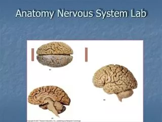
Anatomy Nervous System Lab
Anatomy Nervous System Lab. Divisions of the Nervous System. Central nervous system – brain & spinal cord Peripheral nervous system – cranial nerves & spinal nerves. The Brain. Brain stem medulla oblongata (M.O.) pons midbrain Diencephalon thalamus hypothalamus
954 views • 33 slides

Pathology of Nervous System Basic Anatomy
Pathology of Nervous System Basic Anatomy . VMED 5241 Doo-Youn Cho, DVM, PhD Box 1. Canine Brain (small dog) Dorsal Surface. Identify Structures. Cerebral Hemisphere. Cerebellum. Canine Brain (small dog) Ventral Surface. Identify Structures. Optic Nerve. Pituitary. Oculomotor Nerve.
911 views • 24 slides

Anatomy of the Nervous System
612 views • 55 slides
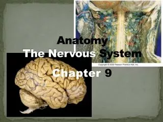
Anatomy The Nervous System
Anatomy The Nervous System. Chapter 9. Nervous system- master controlling & communicating system of body 3 fxns: senses changes within body & in the outside environment (sensory fxn) interprets the changes (integrative fxn)
685 views • 53 slides
The Anatomy of the Nervous System
The Anatomy of the Nervous System. Why study Neuroanatomy? . Major divisions of the nervous system. Major divisions of the nervous system. Brain. Central nervous system. Spinal cord. Afferent nerves. Sensory CNS info. Nervous System. Somatic nervous system.
1.5k views • 57 slides

Anatomy of the Nervous System. Central nervous system (CNS) Brain Spinal cord Peripheral nervous system (PNS) Nerve outside the brain and spinal cord. The 12 pairs of cranial nerves. Spinal Nerves 31 pairs. Spinal Nerves. Dorsal ramus. Ventral ramus.
655 views • 32 slides
Nervous System Anatomy
Nervous System Anatomy. Breakdown:. Central Nervous System (CNS). Peripheral Nervous System (PNS). Somatic Nervous System Autonomic Nervous System Sympathetic N.S. Parasympathetic N.S. Brain Spinal Cord. Function of the CNS:. Spinal Cord Conducts sensory information to the brain
733 views • 46 slides

Central Nervous System Anatomy. Lesson 6. Functional Anatomy: CNS. Major Divisions Forebrain, Midbrain, Hindbrain Know structure name, location & general function Major fiber systems CNS vs PNS terminology nucleus vs. ganglion tracts vs. nerves ~. Telencephalon - Cortex.
365 views • 24 slides
Chapter 4 Anatomy of the Nervous System
Chapter 4 Anatomy of the Nervous System. Structure of the Vertebrate Nervous System. Terms used to describe location when referring to the nervous system include: Ventral Dorsal Anterior Posterior Lateral Medial. More terms…. Lateral Medial Proximal Distal Ipsilateral Contralateral
557 views • 42 slides

NEUROANATOMY Lecture : 12 Anatomy of the Autonomic Nervous System (Involuntary Nervous System)
NEUROANATOMY Lecture : 12 Anatomy of the Autonomic Nervous System (Involuntary Nervous System). Prepared and presented by: Dr. Iyad Mousa Hussein, MD, Ph.D of Neurology Head of Neurology Department Nasser Hospital. LECTURE OBJECTIVES:. Structures of the Autonomic Nervous System.
1.36k views • 93 slides

Comparative Anatomy Nervous System. Witty/Woods PCB Biology. Primary Brain Vesicles. Prosencephalon (Forebrain) Smell Mesoncephalon (Midbrain) Vision Rhombencephalon (Hindbrain) Hearing. Figure 15.1: Primary brain vessicles (book figure 16.13). Primary Brain Vesicles (con’t).
573 views • 29 slides
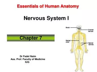
Essentials of Human Anatomy Nervous System I
Essentials of Human Anatomy Nervous System I. Chapter 7. Dr Fadel Naim Ass. Prof. Faculty of Medicine IUG. Introduction. The function of the nervous system, along with the endocrine system, is to communicate The nervous system is made up of the brain, the spinal cord, and the nerves.
561 views • 35 slides

Anatomy of the Central Nervous System: Forebrain. Lecture 5. Telencephalon - Cortex. Gray Matter Somas and Dendrites Neocortex - 6 layers Sensory input ---> layer IV Motor output ---> layer V Paleocortex - 2 or 3 layers ~. Telencephalon - Fiber Systems.
313 views • 18 slides

Chapter 4 Anatomy of the Nervous System. Structure of the Vertebrate Nervous System. Neuroanatomy is the anatomy of the nervous system. Refers to the study of the various parts of the nervous system and their respective function(s).
1.11k views • 90 slides
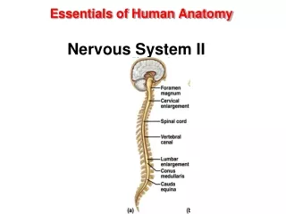
Essentials of Human Anatomy Nervous System II
Essentials of Human Anatomy Nervous System II. Brain. An adult brain weighs between 1.35 and 1.4 kilograms (kg) ( around 3 pounds ) and has a volume of about 1200 cubic centimeters (cc). Brain size is not directly correlated with intelligence
320 views • 28 slides
Got any suggestions?
We want to hear from you! Send us a message and help improve Slidesgo
Top searches
Trending searches

68 templates

33 templates

36 templates

34 templates

9 templates

35 templates
Learn More About the Nervous System Infographics
Free google slides theme and powerpoint template.
The nervous system is responsible for transmitting signals between different parts of the body, allowing us to think, feel, and move. Wow! Take your audience on an exciting journey into the science of the nervous system. These editable infographics for Google Slides & PowerPoint can be a great way of showing data and information about this topic. Besides, you can add them to our template called "Learn More About the Nervous System" if you want to maximize the potential!
Features of these infographics
- 100% editable and easy to modify
- 32 different infographics to boost your presentations
- Include icons and Flaticon’s extension for further customization
- Designed to be used in Google Slides and Microsoft PowerPoint
- 16:9 widescreen format suitable for all types of screens
- Include information about how to edit and customize your infographics
- Supplemental infographics for the template Learn More About the Nervous System
How can I use the infographics?
Am I free to use the templates?
How to attribute the infographics?
Combines with:
This template can be combined with this other one to create the perfect presentation:

Attribution required If you are a free user, you must attribute Slidesgo by keeping the slide where the credits appear. How to attribute?
Related posts on our blog.

How to Add, Duplicate, Move, Delete or Hide Slides in Google Slides

How to Change Layouts in PowerPoint

How to Change the Slide Size in Google Slides
Related presentations.

Premium template
Unlock this template and gain unlimited access


IMAGES
VIDEO
COMMENTS
9. Central Nervous System The central nervous system is composed of two major interconnected organs: - The brain - The spinal cord. These organs work together to integrate and coordinate sensory and motor information for the purpose of controlling the various tissues, organs, and organ systems of the body. The central nervous system is responsible for higher neural functions, such as ...
Free Google Slides theme, PowerPoint template, and Canva presentation template. Discover the fascinating world of the nervous system with this creative template. With calming colors and engaging visuals, you'll be able to explore the different components of the nervous system in a fun and informative way. Plus, you'll get an in-depth look at ...
Central nervous system (CNS) Brain; Spinal cord; Peripheral nervous system (PNS) Nerve outside the brain and spinal cord; 4 of 78. Peripheral Nervous System. ... HTML view of the presentation. Turn on screen reader support To enable screen reader support, press Ctrl+Alt+Z To learn about keyboard shortcuts, press Ctrl+slash ...
1. Electrical signals that are transmitted by the neuron are called neural signals. 2. The junction between 2 neurons is called the Synapse. 3. The transmitting cell is called the sensory neuron. 4. The receiving cell is called the motor neuron. 5.
Cerebrum (cerebral hemispheres) Paired, superior parts of the brain. Includes more than half of the brain mass; obscures most of the brain stem. The surface is made of ridges (gyri = "twisters") and grooves (sulci = "furrows") Fissures (deep grooves) divide the cerebrum into lobes. Occipital lobe. Temporal lobe.
The Nervous System (Slide Show) Nov 24, 2009 •. 1,529 likes • 459,475 views. William Banaag. Lecture about the Nervvous System. Health & Medicine Technology. 1 of 28. The Nervous System (Slide Show) - Download as a PDF or view online for free.
Free Nervous System Slide Templates for an Engaging Slideshow. Make your presentations on the nervous system engaging and informative with a nervous system PowerPoint template. Whether you're a biology teacher, medical student, or neuroscience researcher, these templates will help you convey complex information with clarity and visual appeal.
80. 81. autonomic nervous system regulates the activities of internal organs such as the heart, lungs, blood vessels, digestive organs, and glands. Maintenance and restoration of internal homeostasis is largely the responsibility of the autonomic nervous system. 01-09-2020 81. 82.
Yes! We've risen to the challenge and this is the result: A set of infographics about the nervous system that covers everything from representations of the brain to complete models of humans' nerves. And yes again, they're already labeled, so using them is even easier for you. Have a look at them and if you're students' brains are ...
Template 17: The Neuron: Signals that Matter. The neuron is a specialized cell of the nervous system that is responsible for transmitting signals throughout the body. Use this PPT set to label and explain the structure of the cell and how it functions. Download it now. Download this template.
The Neuron PowerPoint templates for professional presentations are animated diagrams explaining nerve cells' functions and components. The Brain, Neurons, and Nervous System PowerPoint Template include customizable illustrations with descriptions. Users can also change the professional PowerPoint background color or the nervous system ...
Functions of the Nervous System The master controlling & communicating system of the body…. Sensory input —gathering information a. To monitor changes occurring inside and outside the body b. Changes = stimuli. Integration a. To process and interpret sensory input and decide if action is needed. Motor output a.
THE CENTRAL NERVOUS SYSTEM The central nervous system consists of : the Brain and the spinal cord. The Spinal Cord: •is a complex bundle of large nerve fibers protected by bones and spinal fluid. •Transmits messages between the brain and the peripheral nervous system. •Permits some reflex movements.
The professional presentation PowerPoint templates of neurons are animated diagrams to explain the functions and components of nerve cells. The nervous system is the most important biological system in the human body to transmit sensory information. The nervous system has two parts: the central nervous system and the peripheral nervous system.
Free Google Slides theme and PowerPoint template. Download the "Neuroanatomy: Central Nervous System" presentation for PowerPoint or Google Slides. Healthcare goes beyond curing patients and combating illnesses. Raising awareness about diseases, informing people about prevention methods, discussing some good practices, or even talking about a ...
Presentation Transcript. Chapter 22 The Nervous System. Nervous System - Function • Separated into Central and Peripheral Nervous Systems • Receive information about what's happening to the body (both inside & out) • Responds to those internal and environmental stimuli • Maintains homeostasis • Nerve Impulse travels w ...
Nervous system ( anatomy and physiology) Aug 15, 2020 •. 11 likes • 3,014 views. Ravish Yadav. the topic contain function of nervous system, classification of nervous system, neurons anatomy, structural classification of neurons, functional classification of neurons, nerve impulse. Read more. Education. 1 of 43. Download Now.
The Nervous System. Nervous System (597.0K) We welcome your feedback. Please send comments and questions to: [email protected]. To learn more about the book this website supports, please visit its Information Center. ... Home > Chapter 2 > PowerPoint Presentation
Neurology Presentation templates The nervous system is quite complex. The brain, the skin, the muscles, there's not a single part of our body that doesn't play a part! Let's see what the reaction of your audience is when exposed to an stimulus such as that from witnessing these Google Slides themes & PowerPoint templates about neurology.
Download Free and Premium Nervous System PowerPoint Templates. Choose and download Nervous System PowerPoint templates, and Nervous System PowerPoint Backgrounds in just a few minutes.And with amazing ease of use, you can transform your "sleep-inducing" PowerPoint presentation into an aggressive, energetic, jaw-dropping presentation in nearly no time at all.
Functions of the nervous system. 1. Initiate and/or regulate movement of body parts 2. Regulate secretions from glands 3. Gather information about the external environment and the internal environment of the body. Download Presentation. called. nervous system. intermediate zone.
The nervous system is responsible for transmitting signals between different parts of the body, allowing us to think, feel, and move. Wow! Take your audience on an exciting journey into the science of the nervous system. These editable infographics for Google Slides & PowerPoint can be a great way of showing data and information about this ...
The nervous system is a network of neurons whose main feature is to generate, modulate and transmit information between all the different parts of the human body. This property enables many important functions of the nervous system, such as regulation of vital body functions ( heartbeat, breathing, digestion), sensation and body movements.