Head Start Your Radiology Residency [Online] ↗️
- Radiology Thesis – More than 400 Research Topics (2022)!

Please login to bookmark
Introduction
A thesis or dissertation, as some people would like to call it, is an integral part of the Radiology curriculum, be it MD, DNB, or DMRD. We have tried to aggregate radiology thesis topics from various sources for reference.
Not everyone is interested in research, and writing a Radiology thesis can be daunting. But there is no escape from preparing, so it is better that you accept this bitter truth and start working on it instead of cribbing about it (like other things in life. #PhilosophyGyan!)
Start working on your thesis as early as possible and finish your thesis well before your exams, so you do not have that stress at the back of your mind. Also, your thesis may need multiple revisions, so be prepared and allocate time accordingly.
Tips for Choosing Radiology Thesis and Research Topics
Keep it simple silly (kiss).
Retrospective > Prospective
Retrospective studies are better than prospective ones, as you already have the data you need when choosing to do a retrospective study. Prospective studies are better quality, but as a resident, you may not have time (, energy and enthusiasm) to complete these.
Choose a simple topic that answers a single/few questions
Original research is challenging, especially if you do not have prior experience. I would suggest you choose a topic that answers a single or few questions. Most topics that I have listed are along those lines. Alternatively, you can choose a broad topic such as “Role of MRI in evaluation of perianal fistulas.”
You can choose a novel topic if you are genuinely interested in research AND have a good mentor who will guide you. Once you have done that, make sure that you publish your study once you are done with it.
Get it done ASAP.
In most cases, it makes sense to stick to a thesis topic that will not take much time. That does not mean you should ignore your thesis and ‘Ctrl C + Ctrl V’ from a friend from another university. Thesis writing is your first step toward research methodology so do it as sincerely as possible. Do not procrastinate in preparing the thesis. As soon as you have been allotted a guide, start researching topics and writing a review of the literature.
At the same time, do not invest a lot of time in writing/collecting data for your thesis. You should not be busy finishing your thesis a few months before the exam. Some people could not appear for the exam because they could not submit their thesis in time. So DO NOT TAKE thesis lightly.
Do NOT Copy-Paste
Reiterating once again, do not simply choose someone else’s thesis topic. Find out what are kind of cases that your Hospital caters to. It is better to do a good thesis on a common topic than a crappy one on a rare one.
Books to help you write a Radiology Thesis
Event country/university has a different format for thesis; hence these book recommendations may not work for everyone.

- Amazon Kindle Edition
- Gupta, Piyush (Author)
- English (Publication Language)
- 206 Pages - 10/12/2020 (Publication Date) - Jaypee Brothers Medical Publishers (P) Ltd. (Publisher)
In A Hurry? Download a PDF list of Radiology Research Topics!
Sign up below to get this PDF directly to your email address.
100% Privacy Guaranteed. Your information will not be shared. Unsubscribe anytime with a single click.
List of Radiology Research /Thesis / Dissertation Topics
- State of the art of MRI in the diagnosis of hepatic focal lesions
- Multimodality imaging evaluation of sacroiliitis in newly diagnosed patients of spondyloarthropathy
- Multidetector computed tomography in oesophageal varices
- Role of positron emission tomography with computed tomography in the diagnosis of cancer Thyroid
- Evaluation of focal breast lesions using ultrasound elastography
- Role of MRI diffusion tensor imaging in the assessment of traumatic spinal cord injuries
- Sonographic imaging in male infertility
- Comparison of color Doppler and digital subtraction angiography in occlusive arterial disease in patients with lower limb ischemia
- The role of CT urography in Haematuria
- Role of functional magnetic resonance imaging in making brain tumor surgery safer
- Prediction of pre-eclampsia and fetal growth restriction by uterine artery Doppler
- Role of grayscale and color Doppler ultrasonography in the evaluation of neonatal cholestasis
- Validity of MRI in the diagnosis of congenital anorectal anomalies
- Role of sonography in assessment of clubfoot
- Role of diffusion MRI in preoperative evaluation of brain neoplasms
- Imaging of upper airways for pre-anaesthetic evaluation purposes and for laryngeal afflictions.
- A study of multivessel (arterial and venous) Doppler velocimetry in intrauterine growth restriction
- Multiparametric 3tesla MRI of suspected prostatic malignancy.
- Role of Sonography in Characterization of Thyroid Nodules for differentiating benign from
- Role of advances magnetic resonance imaging sequences in multiple sclerosis
- Role of multidetector computed tomography in evaluation of jaw lesions
- Role of Ultrasound and MR Imaging in the Evaluation of Musculotendinous Pathologies of Shoulder Joint
- Role of perfusion computed tomography in the evaluation of cerebral blood flow, blood volume and vascular permeability of cerebral neoplasms
- MRI flow quantification in the assessment of the commonest csf flow abnormalities
- Role of diffusion-weighted MRI in evaluation of prostate lesions and its histopathological correlation
- CT enterography in evaluation of small bowel disorders
- Comparison of perfusion magnetic resonance imaging (PMRI), magnetic resonance spectroscopy (MRS) in and positron emission tomography-computed tomography (PET/CT) in post radiotherapy treated gliomas to detect recurrence
- Role of multidetector computed tomography in evaluation of paediatric retroperitoneal masses
- Role of Multidetector computed tomography in neck lesions
- Estimation of standard liver volume in Indian population
- Role of MRI in evaluation of spinal trauma
- Role of modified sonohysterography in female factor infertility: a pilot study.
- The role of pet-CT in the evaluation of hepatic tumors
- Role of 3D magnetic resonance imaging tractography in assessment of white matter tracts compromise in supratentorial tumors
- Role of dual phase multidetector computed tomography in gallbladder lesions
- Role of multidetector computed tomography in assessing anatomical variants of nasal cavity and paranasal sinuses in patients of chronic rhinosinusitis.
- magnetic resonance spectroscopy in multiple sclerosis
- Evaluation of thyroid nodules by ultrasound elastography using acoustic radiation force impulse (ARFI) imaging
- Role of Magnetic Resonance Imaging in Intractable Epilepsy
- Evaluation of suspected and known coronary artery disease by 128 slice multidetector CT.
- Role of regional diffusion tensor imaging in the evaluation of intracranial gliomas and its histopathological correlation
- Role of chest sonography in diagnosing pneumothorax
- Role of CT virtual cystoscopy in diagnosis of urinary bladder neoplasia
- Role of MRI in assessment of valvular heart diseases
- High resolution computed tomography of temporal bone in unsafe chronic suppurative otitis media
- Multidetector CT urography in the evaluation of hematuria
- Contrast-induced nephropathy in diagnostic imaging investigations with intravenous iodinated contrast media
- Comparison of dynamic susceptibility contrast-enhanced perfusion magnetic resonance imaging and single photon emission computed tomography in patients with little’s disease
- Role of Multidetector Computed Tomography in Bowel Lesions.
- Role of diagnostic imaging modalities in evaluation of post liver transplantation recipient complications.
- Role of multislice CT scan and barium swallow in the estimation of oesophageal tumour length
- Malignant Lesions-A Prospective Study.
- Value of ultrasonography in assessment of acute abdominal diseases in pediatric age group
- Role of three dimensional multidetector CT hysterosalpingography in female factor infertility
- Comparative evaluation of multi-detector computed tomography (MDCT) virtual tracheo-bronchoscopy and fiberoptic tracheo-bronchoscopy in airway diseases
- Role of Multidetector CT in the evaluation of small bowel obstruction
- Sonographic evaluation in adhesive capsulitis of shoulder
- Utility of MR Urography Versus Conventional Techniques in Obstructive Uropathy
- MRI of the postoperative knee
- Role of 64 slice-multi detector computed tomography in diagnosis of bowel and mesenteric injury in blunt abdominal trauma.
- Sonoelastography and triphasic computed tomography in the evaluation of focal liver lesions
- Evaluation of Role of Transperineal Ultrasound and Magnetic Resonance Imaging in Urinary Stress incontinence in Women
- Multidetector computed tomographic features of abdominal hernias
- Evaluation of lesions of major salivary glands using ultrasound elastography
- Transvaginal ultrasound and magnetic resonance imaging in female urinary incontinence
- MDCT colonography and double-contrast barium enema in evaluation of colonic lesions
- Role of MRI in diagnosis and staging of urinary bladder carcinoma
- Spectrum of imaging findings in children with febrile neutropenia.
- Spectrum of radiographic appearances in children with chest tuberculosis.
- Role of computerized tomography in evaluation of mediastinal masses in pediatric
- Diagnosing renal artery stenosis: Comparison of multimodality imaging in diabetic patients
- Role of multidetector CT virtual hysteroscopy in the detection of the uterine & tubal causes of female infertility
- Role of multislice computed tomography in evaluation of crohn’s disease
- CT quantification of parenchymal and airway parameters on 64 slice MDCT in patients of chronic obstructive pulmonary disease
- Comparative evaluation of MDCT and 3t MRI in radiographically detected jaw lesions.
- Evaluation of diagnostic accuracy of ultrasonography, colour Doppler sonography and low dose computed tomography in acute appendicitis
- Ultrasonography , magnetic resonance cholangio-pancreatography (MRCP) in assessment of pediatric biliary lesions
- Multidetector computed tomography in hepatobiliary lesions.
- Evaluation of peripheral nerve lesions with high resolution ultrasonography and colour Doppler
- Multidetector computed tomography in pancreatic lesions
- Multidetector Computed Tomography in Paediatric abdominal masses.
- Evaluation of focal liver lesions by colour Doppler and MDCT perfusion imaging
- Sonographic evaluation of clubfoot correction during Ponseti treatment
- Role of multidetector CT in characterization of renal masses
- Study to assess the role of Doppler ultrasound in evaluation of arteriovenous (av) hemodialysis fistula and the complications of hemodialysis vasular access
- Comparative study of multiphasic contrast-enhanced CT and contrast-enhanced MRI in the evaluation of hepatic mass lesions
- Sonographic spectrum of rheumatoid arthritis
- Diagnosis & staging of liver fibrosis by ultrasound elastography in patients with chronic liver diseases
- Role of multidetector computed tomography in assessment of jaw lesions.
- Role of high-resolution ultrasonography in the differentiation of benign and malignant thyroid lesions
- Radiological evaluation of aortic aneurysms in patients selected for endovascular repair
- Role of conventional MRI, and diffusion tensor imaging tractography in evaluation of congenital brain malformations
- To evaluate the status of coronary arteries in patients with non-valvular atrial fibrillation using 256 multirow detector CT scan
- A comparative study of ultrasonography and CT – arthrography in diagnosis of chronic ligamentous and meniscal injuries of knee
- Multi detector computed tomography evaluation in chronic obstructive pulmonary disease and correlation with severity of disease
- Diffusion weighted and dynamic contrast enhanced magnetic resonance imaging in chemoradiotherapeutic response evaluation in cervical cancer.
- High resolution sonography in the evaluation of non-traumatic painful wrist
- The role of trans-vaginal ultrasound versus magnetic resonance imaging in diagnosis & evaluation of cancer cervix
- Role of multidetector row computed tomography in assessment of maxillofacial trauma
- Imaging of vascular complication after liver transplantation.
- Role of magnetic resonance perfusion weighted imaging & spectroscopy for grading of glioma by correlating perfusion parameter of the lesion with the final histopathological grade
- Magnetic resonance evaluation of abdominal tuberculosis.
- Diagnostic usefulness of low dose spiral HRCT in diffuse lung diseases
- Role of dynamic contrast enhanced and diffusion weighted magnetic resonance imaging in evaluation of endometrial lesions
- Contrast enhanced digital mammography anddigital breast tomosynthesis in early diagnosis of breast lesion
- Evaluation of Portal Hypertension with Colour Doppler flow imaging and magnetic resonance imaging
- Evaluation of musculoskeletal lesions by magnetic resonance imaging
- Role of diffusion magnetic resonance imaging in assessment of neoplastic and inflammatory brain lesions
- Radiological spectrum of chest diseases in HIV infected children High resolution ultrasonography in neck masses in children
- with surgical findings
- Sonographic evaluation of peripheral nerves in type 2 diabetes mellitus.
- Role of perfusion computed tomography in the evaluation of neck masses and correlation
- Role of ultrasonography in the diagnosis of knee joint lesions
- Role of ultrasonography in evaluation of various causes of pelvic pain in first trimester of pregnancy.
- Role of Magnetic Resonance Angiography in the Evaluation of Diseases of Aorta and its Branches
- MDCT fistulography in evaluation of fistula in Ano
- Role of multislice CT in diagnosis of small intestine tumors
- Role of high resolution CT in differentiation between benign and malignant pulmonary nodules in children
- A study of multidetector computed tomography urography in urinary tract abnormalities
- Role of high resolution sonography in assessment of ulnar nerve in patients with leprosy.
- Pre-operative radiological evaluation of locally aggressive and malignant musculoskeletal tumours by computed tomography and magnetic resonance imaging.
- The role of ultrasound & MRI in acute pelvic inflammatory disease
- Ultrasonography compared to computed tomographic arthrography in the evaluation of shoulder pain
- Role of Multidetector Computed Tomography in patients with blunt abdominal trauma.
- The Role of Extended field-of-view Sonography and compound imaging in Evaluation of Breast Lesions
- Evaluation of focal pancreatic lesions by Multidetector CT and perfusion CT
- Evaluation of breast masses on sono-mammography and colour Doppler imaging
- Role of CT virtual laryngoscopy in evaluation of laryngeal masses
- Triple phase multi detector computed tomography in hepatic masses
- Role of transvaginal ultrasound in diagnosis and treatment of female infertility
- Role of ultrasound and color Doppler imaging in assessment of acute abdomen due to female genetal causes
- High resolution ultrasonography and color Doppler ultrasonography in scrotal lesion
- Evaluation of diagnostic accuracy of ultrasonography with colour Doppler vs low dose computed tomography in salivary gland disease
- Role of multidetector CT in diagnosis of salivary gland lesions
- Comparison of diagnostic efficacy of ultrasonography and magnetic resonance cholangiopancreatography in obstructive jaundice: A prospective study
- Evaluation of varicose veins-comparative assessment of low dose CT venogram with sonography: pilot study
- Role of mammotome in breast lesions
- The role of interventional imaging procedures in the treatment of selected gynecological disorders
- Role of transcranial ultrasound in diagnosis of neonatal brain insults
- Role of multidetector CT virtual laryngoscopy in evaluation of laryngeal mass lesions
- Evaluation of adnexal masses on sonomorphology and color Doppler imaginig
- Role of radiological imaging in diagnosis of endometrial carcinoma
- Comprehensive imaging of renal masses by magnetic resonance imaging
- The role of 3D & 4D ultrasonography in abnormalities of fetal abdomen
- Diffusion weighted magnetic resonance imaging in diagnosis and characterization of brain tumors in correlation with conventional MRI
- Role of diffusion weighted MRI imaging in evaluation of cancer prostate
- Role of multidetector CT in diagnosis of urinary bladder cancer
- Role of multidetector computed tomography in the evaluation of paediatric retroperitoneal masses.
- Comparative evaluation of gastric lesions by double contrast barium upper G.I. and multi detector computed tomography
- Evaluation of hepatic fibrosis in chronic liver disease using ultrasound elastography
- Role of MRI in assessment of hydrocephalus in pediatric patients
- The role of sonoelastography in characterization of breast lesions
- The influence of volumetric tumor doubling time on survival of patients with intracranial tumours
- Role of perfusion computed tomography in characterization of colonic lesions
- Role of proton MRI spectroscopy in the evaluation of temporal lobe epilepsy
- Role of Doppler ultrasound and multidetector CT angiography in evaluation of peripheral arterial diseases.
- Role of multidetector computed tomography in paranasal sinus pathologies
- Role of virtual endoscopy using MDCT in detection & evaluation of gastric pathologies
- High resolution 3 Tesla MRI in the evaluation of ankle and hindfoot pain.
- Transperineal ultrasonography in infants with anorectal malformation
- CT portography using MDCT versus color Doppler in detection of varices in cirrhotic patients
- Role of CT urography in the evaluation of a dilated ureter
- Characterization of pulmonary nodules by dynamic contrast-enhanced multidetector CT
- Comprehensive imaging of acute ischemic stroke on multidetector CT
- The role of fetal MRI in the diagnosis of intrauterine neurological congenital anomalies
- Role of Multidetector computed tomography in pediatric chest masses
- Multimodality imaging in the evaluation of palpable & non-palpable breast lesion.
- Sonographic Assessment Of Fetal Nasal Bone Length At 11-28 Gestational Weeks And Its Correlation With Fetal Outcome.
- Role Of Sonoelastography And Contrast-Enhanced Computed Tomography In Evaluation Of Lymph Node Metastasis In Head And Neck Cancers
- Role Of Renal Doppler And Shear Wave Elastography In Diabetic Nephropathy
- Evaluation Of Relationship Between Various Grades Of Fatty Liver And Shear Wave Elastography Values
- Evaluation and characterization of pelvic masses of gynecological origin by USG, color Doppler and MRI in females of reproductive age group
- Radiological evaluation of small bowel diseases using computed tomographic enterography
- Role of coronary CT angiography in patients of coronary artery disease
- Role of multimodality imaging in the evaluation of pediatric neck masses
- Role of CT in the evaluation of craniocerebral trauma
- Role of magnetic resonance imaging (MRI) in the evaluation of spinal dysraphism
- Comparative evaluation of triple phase CT and dynamic contrast-enhanced MRI in patients with liver cirrhosis
- Evaluation of the relationship between carotid intima-media thickness and coronary artery disease in patients evaluated by coronary angiography for suspected CAD
- Assessment of hepatic fat content in fatty liver disease by unenhanced computed tomography
- Correlation of vertebral marrow fat on spectroscopy and diffusion-weighted MRI imaging with bone mineral density in postmenopausal women.
- Comparative evaluation of CT coronary angiography with conventional catheter coronary angiography
- Ultrasound evaluation of kidney length & descending colon diameter in normal and intrauterine growth-restricted fetuses
- A prospective study of hepatic vein waveform and splenoportal index in liver cirrhosis: correlation with child Pugh’s classification and presence of esophageal varices.
- CT angiography to evaluate coronary artery by-pass graft patency in symptomatic patient’s functional assessment of myocardium by cardiac MRI in patients with myocardial infarction
- MRI evaluation of HIV positive patients with central nervous system manifestations
- MDCT evaluation of mediastinal and hilar masses
- Evaluation of rotator cuff & labro-ligamentous complex lesions by MRI & MRI arthrography of shoulder joint
- Role of imaging in the evaluation of soft tissue vascular malformation
- Role of MRI and ultrasonography in the evaluation of multifidus muscle pathology in chronic low back pain patients
- Role of ultrasound elastography in the differential diagnosis of breast lesions
- Role of magnetic resonance cholangiopancreatography in evaluating dilated common bile duct in patients with symptomatic gallstone disease.
- Comparative study of CT urography & hybrid CT urography in patients with haematuria.
- Role of MRI in the evaluation of anorectal malformations
- Comparison of ultrasound-Doppler and magnetic resonance imaging findings in rheumatoid arthritis of hand and wrist
- Role of Doppler sonography in the evaluation of renal artery stenosis in hypertensive patients undergoing coronary angiography for coronary artery disease.
- Comparison of radiography, computed tomography and magnetic resonance imaging in the detection of sacroiliitis in ankylosing spondylitis.
- Mr evaluation of painful hip
- Role of MRI imaging in pretherapeutic assessment of oral and oropharyngeal malignancy
- Evaluation of diffuse lung diseases by high resolution computed tomography of the chest
- Mr evaluation of brain parenchyma in patients with craniosynostosis.
- Diagnostic and prognostic value of cardiovascular magnetic resonance imaging in dilated cardiomyopathy
- Role of multiparametric magnetic resonance imaging in the detection of early carcinoma prostate
- Role of magnetic resonance imaging in white matter diseases
- Role of sonoelastography in assessing the response to neoadjuvant chemotherapy in patients with locally advanced breast cancer.
- Role of ultrasonography in the evaluation of carotid and femoral intima-media thickness in predialysis patients with chronic kidney disease
- Role of H1 MRI spectroscopy in focal bone lesions of peripheral skeleton choline detection by MRI spectroscopy in breast cancer and its correlation with biomarkers and histological grade.
- Ultrasound and MRI evaluation of axillary lymph node status in breast cancer.
- Role of sonography and magnetic resonance imaging in evaluating chronic lateral epicondylitis.
- Comparative of sonography including Doppler and sonoelastography in cervical lymphadenopathy.
- Evaluation of Umbilical Coiling Index as Predictor of Pregnancy Outcome.
- Computerized Tomographic Evaluation of Azygoesophageal Recess in Adults.
- Lumbar Facet Arthropathy in Low Backache.
- “Urethral Injuries After Pelvic Trauma: Evaluation with Uretrography
- Role Of Ct In Diagnosis Of Inflammatory Renal Diseases
- Role Of Ct Virtual Laryngoscopy In Evaluation Of Laryngeal Masses
- “Ct Portography Using Mdct Versus Color Doppler In Detection Of Varices In
- Cirrhotic Patients”
- Role Of Multidetector Ct In Characterization Of Renal Masses
- Role Of Ct Virtual Cystoscopy In Diagnosis Of Urinary Bladder Neoplasia
- Role Of Multislice Ct In Diagnosis Of Small Intestine Tumors
- “Mri Flow Quantification In The Assessment Of The Commonest CSF Flow Abnormalities”
- “The Role Of Fetal Mri In Diagnosis Of Intrauterine Neurological CongenitalAnomalies”
- Role Of Transcranial Ultrasound In Diagnosis Of Neonatal Brain Insults
- “The Role Of Interventional Imaging Procedures In The Treatment Of Selected Gynecological Disorders”
- Role Of Radiological Imaging In Diagnosis Of Endometrial Carcinoma
- “Role Of High-Resolution Ct In Differentiation Between Benign And Malignant Pulmonary Nodules In Children”
- Role Of Ultrasonography In The Diagnosis Of Knee Joint Lesions
- “Role Of Diagnostic Imaging Modalities In Evaluation Of Post Liver Transplantation Recipient Complications”
- “Diffusion-Weighted Magnetic Resonance Imaging In Diagnosis And
- Characterization Of Brain Tumors In Correlation With Conventional Mri”
- The Role Of PET-CT In The Evaluation Of Hepatic Tumors
- “Role Of Computerized Tomography In Evaluation Of Mediastinal Masses In Pediatric patients”
- “Trans Vaginal Ultrasound And Magnetic Resonance Imaging In Female Urinary Incontinence”
- Role Of Multidetector Ct In Diagnosis Of Urinary Bladder Cancer
- “Role Of Transvaginal Ultrasound In Diagnosis And Treatment Of Female Infertility”
- Role Of Diffusion-Weighted Mri Imaging In Evaluation Of Cancer Prostate
- “Role Of Positron Emission Tomography With Computed Tomography In Diagnosis Of Cancer Thyroid”
- The Role Of CT Urography In Case Of Haematuria
- “Value Of Ultrasonography In Assessment Of Acute Abdominal Diseases In Pediatric Age Group”
- “Role Of Functional Magnetic Resonance Imaging In Making Brain Tumor Surgery Safer”
- The Role Of Sonoelastography In Characterization Of Breast Lesions
- “Ultrasonography, Magnetic Resonance Cholangiopancreatography (MRCP) In Assessment Of Pediatric Biliary Lesions”
- “Role Of Ultrasound And Color Doppler Imaging In Assessment Of Acute Abdomen Due To Female Genital Causes”
- “Role Of Multidetector Ct Virtual Laryngoscopy In Evaluation Of Laryngeal Mass Lesions”
- MRI Of The Postoperative Knee
- Role Of Mri In Assessment Of Valvular Heart Diseases
- The Role Of 3D & 4D Ultrasonography In Abnormalities Of Fetal Abdomen
- State Of The Art Of Mri In Diagnosis Of Hepatic Focal Lesions
- Role Of Multidetector Ct In Diagnosis Of Salivary Gland Lesions
- “Role Of Virtual Endoscopy Using Mdct In Detection & Evaluation Of Gastric Pathologies”
- The Role Of Ultrasound & Mri In Acute Pelvic Inflammatory Disease
- “Diagnosis & Staging Of Liver Fibrosis By Ultraso Und Elastography In
- Patients With Chronic Liver Diseases”
- Role Of Mri In Evaluation Of Spinal Trauma
- Validity Of Mri In Diagnosis Of Congenital Anorectal Anomalies
- Imaging Of Vascular Complication After Liver Transplantation
- “Contrast-Enhanced Digital Mammography And Digital Breast Tomosynthesis In Early Diagnosis Of Breast Lesion”
- Role Of Mammotome In Breast Lesions
- “Role Of MRI Diffusion Tensor Imaging (DTI) In Assessment Of Traumatic Spinal Cord Injuries”
- “Prediction Of Pre-eclampsia And Fetal Growth Restriction By Uterine Artery Doppler”
- “Role Of Multidetector Row Computed Tomography In Assessment Of Maxillofacial Trauma”
- “Role Of Diffusion Magnetic Resonance Imaging In Assessment Of Neoplastic And Inflammatory Brain Lesions”
- Role Of Diffusion Mri In Preoperative Evaluation Of Brain Neoplasms
- “Role Of Multidetector Ct Virtual Hysteroscopy In The Detection Of The
- Uterine & Tubal Causes Of Female Infertility”
- Role Of Advances Magnetic Resonance Imaging Sequences In Multiple Sclerosis Magnetic Resonance Spectroscopy In Multiple Sclerosis
- “Role Of Conventional Mri, And Diffusion Tensor Imaging Tractography In Evaluation Of Congenital Brain Malformations”
- Role Of MRI In Evaluation Of Spinal Trauma
- Diagnostic Role Of Diffusion-weighted MR Imaging In Neck Masses
- “The Role Of Transvaginal Ultrasound Versus Magnetic Resonance Imaging In Diagnosis & Evaluation Of Cancer Cervix”
- “Role Of 3d Magnetic Resonance Imaging Tractography In Assessment Of White Matter Tracts Compromise In Supra Tentorial Tumors”
- Role Of Proton MR Spectroscopy In The Evaluation Of Temporal Lobe Epilepsy
- Role Of Multislice Computed Tomography In Evaluation Of Crohn’s Disease
- Role Of MRI In Assessment Of Hydrocephalus In Pediatric Patients
- The Role Of MRI In Diagnosis And Staging Of Urinary Bladder Carcinoma
- USG and MRI correlation of congenital CNS anomalies
- HRCT in interstitial lung disease
- X-Ray, CT and MRI correlation of bone tumors
- “Study on the diagnostic and prognostic utility of X-Rays for cases of pulmonary tuberculosis under RNTCP”
- “Role of magnetic resonance imaging in the characterization of female adnexal pathology”
- “CT angiography of carotid atherosclerosis and NECT brain in cerebral ischemia, a correlative analysis”
- Role of CT scan in the evaluation of paranasal sinus pathology
- USG and MRI correlation on shoulder joint pathology
- “Radiological evaluation of a patient presenting with extrapulmonary tuberculosis”
- CT and MRI correlation in focal liver lesions”
- Comparison of MDCT virtual cystoscopy with conventional cystoscopy in bladder tumors”
- “Bleeding vessels in life-threatening hemoptysis: Comparison of 64 detector row CT angiography with conventional angiography prior to endovascular management”
- “Role of transarterial chemoembolization in unresectable hepatocellular carcinoma”
- “Comparison of color flow duplex study with digital subtraction angiography in the evaluation of peripheral vascular disease”
- “A Study to assess the efficacy of magnetization transfer ratio in differentiating tuberculoma from neurocysticercosis”
- “MR evaluation of uterine mass lesions in correlation with transabdominal, transvaginal ultrasound using HPE as a gold standard”
- “The Role of power Doppler imaging with trans rectal ultrasonogram guided prostate biopsy in the detection of prostate cancer”
- “Lower limb arteries assessed with doppler angiography – A prospective comparative study with multidetector CT angiography”
- “Comparison of sildenafil with papaverine in penile doppler by assessing hemodynamic changes”
- “Evaluation of efficacy of sonosalphingogram for assessing tubal patency in infertile patients with hysterosalpingogram as the gold standard”
- Role of CT enteroclysis in the evaluation of small bowel diseases
- “MRI colonography versus conventional colonoscopy in the detection of colonic polyposis”
- “Magnetic Resonance Imaging of anteroposterior diameter of the midbrain – differentiation of progressive supranuclear palsy from Parkinson disease”
- “MRI Evaluation of anterior cruciate ligament tears with arthroscopic correlation”
- “The Clinicoradiological profile of cerebral venous sinus thrombosis with prognostic evaluation using MR sequences”
- “Role of MRI in the evaluation of pelvic floor integrity in stress incontinent patients” “Doppler ultrasound evaluation of hepatic venous waveform in portal hypertension before and after propranolol”
- “Role of transrectal sonography with colour doppler and MRI in evaluation of prostatic lesions with TRUS guided biopsy correlation”
- “Ultrasonographic evaluation of painful shoulders and correlation of rotator cuff pathologies and clinical examination”
- “Colour Doppler Evaluation of Common Adult Hepatic tumors More Than 2 Cm with HPE and CECT Correlation”
- “Clinical Relevance of MR Urethrography in Obliterative Posterior Urethral Stricture”
- “Prediction of Adverse Perinatal Outcome in Growth Restricted Fetuses with Antenatal Doppler Study”
- Radiological evaluation of spinal dysraphism using CT and MRI
- “Evaluation of temporal bone in cholesteatoma patients by high resolution computed tomography”
- “Radiological evaluation of primary brain tumours using computed tomography and magnetic resonance imaging”
- “Three dimensional colour doppler sonographic assessment of changes in volume and vascularity of fibroids – before and after uterine artery embolization”
- “In phase opposed phase imaging of bone marrow differentiating neoplastic lesions”
- “Role of dynamic MRI in replacing the isotope renogram in the functional evaluation of PUJ obstruction”
- Characterization of adrenal masses with contrast-enhanced CT – washout study
- A study on accuracy of magnetic resonance cholangiopancreatography
- “Evaluation of median nerve in carpal tunnel syndrome by high-frequency ultrasound & color doppler in comparison with nerve conduction studies”
- “Correlation of Agatston score in patients with obstructive and nonobstructive coronary artery disease following STEMI”
- “Doppler ultrasound assessment of tumor vascularity in locally advanced breast cancer at diagnosis and following primary systemic chemotherapy.”
- “Validation of two-dimensional perineal ultrasound and dynamic magnetic resonance imaging in pelvic floor dysfunction.”
- “Role of MR urethrography compared to conventional urethrography in the surgical management of obliterative urethral stricture.”
Search Diagnostic Imaging Research Topics
You can also search research-related resources on our custom search engine .
Free Resources for Preparing Radiology Thesis
- Radiology thesis topics- Benha University – Free to download thesis
- Radiology thesis topics – Faculty of Medical Science Delhi
- Radiology thesis topics – IPGMER
- Fetal Radiology thesis Protocols
- Radiology thesis and dissertation topics
- Radiographics
Proofreading Your Thesis:
Make sure you use Grammarly to correct your spelling , grammar , and plagiarism for your thesis. Grammarly has affordable paid subscriptions, windows/macOS apps, and FREE browser extensions. It is an excellent tool to avoid inadvertent spelling mistakes in your research projects. It has an extensive built-in vocabulary, but you should make an account and add your own medical glossary to it.

Guidelines for Writing a Radiology Thesis:
These are general guidelines and not about radiology specifically. You can share these with colleagues from other departments as well. Special thanks to Dr. Sanjay Yadav sir for these. This section is best seen on a desktop. Here are a couple of handy presentations to start writing a thesis:
Read the general guidelines for writing a thesis (the page will take some time to load- more than 70 pages!
A format for thesis protocol with a sample patient information sheet, sample patient consent form, sample application letter for thesis, and sample certificate.
Resources and References:
- Guidelines for thesis writing.
- Format for thesis protocol
- Thesis protocol writing guidelines DNB
- Informed consent form for Research studies from AIIMS
- Radiology Informed consent forms in local Indian languages.
- Sample Informed Consent form for Research in Hindi
- Guide to write a thesis by Dr. P R Sharma
- Guidelines for thesis writing by Dr. Pulin Gupta.
- Preparing MD/DNB thesis by A Indrayan
- Another good thesis reference protocol
Hopefully, this post will make the tedious task of writing a Radiology thesis a little bit easier for you. Best of luck with writing your thesis and your residency too!
More guides for residents :
- Guide for the MD/DMRD/DNB radiology exam!
- Guide for First-Year Radiology Residents
- FRCR Exam: THE Most Comprehensive Guide (2022)!
- Radiology Practical Exams Questions compilation for MD/DNB/DMRD !
Radiology Exam Resources (Oral Recalls, Instruments, etc )!
- Tips and Tricks for DNB/MD Radiology Practical Exam
- FRCR 2B exam- Tips and Tricks !
FRCR exam preparation – An alternative take!
- Why did I take up Radiology?
- Radiology Conferences – A comprehensive guide!
- ECR (European Congress Of Radiology)
- European Diploma in Radiology (EDiR) – The Complete Guide!
- Radiology NEET PG guide – How to select THE best college for post-graduation in Radiology (includes personal insights)!
- Interventional Radiology – All Your Questions Answered!
- What It Means To Be A Radiologist: A Guide For Medical Students!
- Radiology Mentors for Medical Students (Post NEET-PG)
- MD vs DNB Radiology: Which Path is Right for Your Career?
- DNB Radiology OSCE – Tips and Tricks
More radiology resources here: Radiology resources This page will be updated regularly. Kindly leave your feedback in the comments or send us a message here . Also, you can comment below regarding your department’s thesis topics.
Note: All topics have been compiled from available online resources. If anyone has an issue with any radiology thesis topics displayed here, you can message us here , and we can delete them. These are only sample guidelines. Thesis guidelines differ from institution to institution.
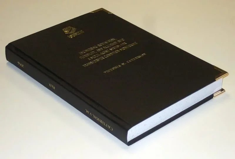
Image source: Thesis complete! (2018). Flickr. Retrieved 12 August 2018, from https://www.flickr.com/photos/cowlet/354911838 by Victoria Catterson
About The Author
Dr. amar udare, md, related posts ↓.
7 thoughts on “Radiology Thesis – More than 400 Research Topics (2022)!”
Amazing & The most helpful site for Radiology residents…
Thank you for your kind comments 🙂
Dr. I saw your Tips is very amazing and referable. But Dr. Can you help me with the thesis of Evaluation of Diagnostic accuracy of X-ray radiograph in knee joint lesion.
Wow! These are excellent stuff. You are indeed a teacher. God bless
Glad you liked these!
happy to see this
Glad I could help :).
Leave a Comment Cancel Reply
Your email address will not be published. Required fields are marked *
Get Radiology Updates to Your Inbox!
This site is for use by medical professionals. To continue, you must accept our use of cookies and the site's Terms of Use. Learn more Accept!
Wish to be a BETTER Radiologist? Join 14000 Radiology Colleagues !
Enter your email address below to access HIGH YIELD radiology content, updates, and resources.
No spam, only VALUE! Unsubscribe anytime with a single click.
Radiology Research Paper Topics

Radiology research paper topics encompass a wide range of fascinating areas within the field of medical imaging. This page aims to provide students studying health sciences with a comprehensive collection of radiology research paper topics to inspire and guide their research endeavors. By delving into various categories and exploring ten thought-provoking topics within each, students can gain insights into the diverse research possibilities in radiology. From advancements in imaging technology to the evaluation of diagnostic accuracy and the impact of radiological interventions, these topics offer a glimpse into the exciting world of radiology research. Additionally, expert advice is provided to help students choose the most suitable research topics and navigate the process of writing a research paper in radiology. By leveraging iResearchNet’s writing services, students can further enhance their research papers with professional assistance, ensuring the highest quality and adherence to academic standards. Explore the realm of radiology research paper topics and unleash your potential to contribute to the advancement of medical imaging and patient care.
100 Radiology Research Paper Topics
Radiology encompasses a broad spectrum of imaging techniques used to diagnose diseases, monitor treatment progress, and guide interventions. This comprehensive list of radiology research paper topics serves as a valuable resource for students in the field of health sciences who are seeking inspiration and guidance for their research endeavors. The following ten categories highlight different areas within radiology, each containing ten thought-provoking topics. Exploring these topics will provide students with a deeper understanding of the diverse research possibilities and current trends within the field of radiology.
Academic Writing, Editing, Proofreading, And Problem Solving Services
Get 10% off with 24start discount code.
Diagnostic Imaging Techniques
- Comparative analysis of imaging modalities: CT, MRI, and PET-CT.
- The role of artificial intelligence in radiological image interpretation.
- Advancements in digital mammography for breast cancer screening.
- Emerging techniques in nuclear medicine imaging.
- Image-guided biopsy: Enhancing accuracy and safety.
- Application of radiomics in predicting treatment response.
- Dual-energy CT: Expanding diagnostic capabilities.
- Radiological evaluation of traumatic brain injuries.
- Imaging techniques for evaluating cardiovascular diseases.
- Radiographic evaluation of pulmonary nodules: Challenges and advancements.
Interventional Radiology
- Minimally invasive treatments for liver tumors: Embolization techniques.
- Radiofrequency ablation in the management of renal cell carcinoma.
- Role of interventional radiology in the treatment of peripheral artery disease.
- Transarterial chemoembolization in hepatocellular carcinoma.
- Evaluation of uterine artery embolization for the treatment of fibroids.
- Percutaneous vertebroplasty and kyphoplasty: Efficacy and complications.
- Endovascular repair of abdominal aortic aneurysms: Long-term outcomes.
- Interventional radiology in the management of deep vein thrombosis.
- Transcatheter aortic valve replacement: Imaging considerations.
- Emerging techniques in interventional oncology.
Radiation Safety and Dose Optimization
- Strategies for reducing radiation dose in pediatric imaging.
- Imaging modalities with low radiation exposure: Current advancements.
- Effective use of dose monitoring systems in radiology departments.
- The impact of artificial intelligence on radiation dose optimization.
- Optimization of radiation therapy treatment plans: Balancing efficacy and safety.
- Radioprotective measures for patients and healthcare professionals.
- The role of radiology in addressing radiation-induced risks.
- Evaluating the long-term effects of radiation exposure in diagnostic imaging.
- Radiation dose tracking and reporting: Implementing best practices.
- Patient education and communication regarding radiation risks.
Radiology in Oncology
- Imaging techniques for early detection and staging of lung cancer.
- Quantitative imaging biomarkers for predicting treatment response in solid tumors.
- Radiogenomics: Linking imaging features to genetic profiles in cancer.
- The role of imaging in assessing tumor angiogenesis.
- Radiological evaluation of lymphoma: Challenges and advancements.
- Imaging-guided interventions in the treatment of hepatocellular carcinoma.
- Assessment of tumor heterogeneity using functional imaging techniques.
- Radiomics and machine learning in predicting treatment outcomes in cancer.
- Multimodal imaging in the evaluation of brain tumors.
- Imaging surveillance after cancer treatment: Optimizing follow-up protocols.
Radiology in Musculoskeletal Disorders
- Imaging modalities in the evaluation of sports-related injuries.
- The role of imaging in diagnosing and monitoring rheumatoid arthritis.
- Assessment of bone health using dual-energy X-ray absorptiometry (DXA).
- Imaging techniques for evaluating osteoarthritis progression.
- Imaging-guided interventions in the management of musculoskeletal tumors.
- Role of imaging in diagnosing and managing spinal disorders.
- Evaluation of traumatic injuries using radiography, CT, and MRI.
- Imaging of joint prostheses: Complications and assessment techniques.
- Imaging features and classifications of bone fractures.
- Musculoskeletal ultrasound in the diagnosis of soft tissue injuries.
Neuroradiology
- Advanced neuroimaging techniques for early detection of neurodegenerative diseases.
- Imaging evaluation of acute stroke: Current guidelines and advancements.
- Role of functional MRI in mapping brain functions.
- Imaging of brain tumors: Classification and treatment planning.
- Diffusion tensor imaging in assessing white matter integrity.
- Neuroimaging in the evaluation of multiple sclerosis.
- Imaging techniques for the assessment of epilepsy.
- Radiological evaluation of neurovascular diseases.
- Imaging of cranial nerve disorders: Diagnosis and management.
- Radiological assessment of developmental brain abnormalities.
Pediatric Radiology
- Radiation dose reduction strategies in pediatric imaging.
- Imaging evaluation of congenital heart diseases in children.
- Role of imaging in the diagnosis and management of pediatric oncology.
- Imaging of pediatric gastrointestinal disorders.
- Evaluation of developmental hip dysplasia using ultrasound and radiography.
- Imaging features and management of pediatric musculoskeletal infections.
- Neuroimaging in the assessment of pediatric neurodevelopmental disorders.
- Radiological evaluation of pediatric respiratory conditions.
- Imaging techniques for the evaluation of pediatric abdominal emergencies.
- Imaging-guided interventions in pediatric patients.
Breast Imaging
- Advances in digital mammography for early breast cancer detection.
- The role of tomosynthesis in breast imaging.
- Imaging evaluation of breast implants: Complications and assessment.
- Radiogenomic analysis of breast cancer subtypes.
- Contrast-enhanced mammography: Diagnostic benefits and challenges.
- Emerging techniques in breast MRI for high-risk populations.
- Evaluation of breast density and its implications for cancer risk.
- Role of molecular breast imaging in dense breast tissue evaluation.
- Radiological evaluation of male breast disorders.
- The impact of artificial intelligence on breast cancer screening.
Cardiac Imaging
- Imaging evaluation of coronary artery disease: Current techniques and challenges.
- Role of cardiac CT angiography in the assessment of structural heart diseases.
- Imaging of cardiac tumors: Diagnosis and treatment considerations.
- Advanced imaging techniques for assessing myocardial viability.
- Evaluation of valvular heart diseases using echocardiography and MRI.
- Cardiac magnetic resonance imaging in the evaluation of cardiomyopathies.
- Role of nuclear cardiology in the assessment of cardiac function.
- Imaging evaluation of congenital heart diseases in adults.
- Radiological assessment of cardiac arrhythmias.
- Imaging-guided interventions in structural heart diseases.
Abdominal and Pelvic Imaging
- Evaluation of hepatobiliary diseases using imaging techniques.
- Imaging features and classification of renal masses.
- Radiological assessment of gastrointestinal bleeding.
- Imaging evaluation of pancreatic diseases: Challenges and advancements.
- Evaluation of pelvic floor disorders using MRI and ultrasound.
- Role of imaging in diagnosing and staging gynecological cancers.
- Imaging of abdominal and pelvic trauma: Current guidelines and techniques.
- Radiological evaluation of genitourinary disorders.
- Imaging features of abdominal and pelvic infections.
- Assessment of abdominal and pelvic vascular diseases using imaging techniques.
This comprehensive list of radiology research paper topics highlights the vast range of research possibilities within the field of medical imaging. Each category offers unique insights and avenues for exploration, enabling students to delve into various aspects of radiology. By choosing a topic of interest and relevance, students can contribute to the advancement of medical imaging and patient care. The provided topics serve as a starting point for students to engage in in-depth research and produce high-quality research papers.
Radiology: Exploring the Range of Research Paper Topics
Introduction: Radiology plays a crucial role in modern healthcare, providing valuable insights into the diagnosis, treatment, and monitoring of various medical conditions. As a dynamic and rapidly evolving field, radiology offers a wide range of research opportunities for students in the health sciences. This article aims to explore the diverse spectrum of research paper topics within radiology, shedding light on the current trends, innovations, and challenges in the field.
Radiology in Diagnostic Imaging : Diagnostic imaging is one of the core areas of radiology, encompassing various modalities such as X-ray, computed tomography (CT), magnetic resonance imaging (MRI), ultrasound, and nuclear medicine. Research topics in this domain may include advancements in imaging techniques, comparative analysis of modalities, radiomics, and the integration of artificial intelligence in image interpretation. Students can explore how these technological advancements enhance diagnostic accuracy, improve patient outcomes, and optimize radiation exposure.
Interventional Radiology : Interventional radiology focuses on minimally invasive procedures performed under image guidance. Research topics in this area can cover a wide range of interventions, such as angioplasty, embolization, radiofrequency ablation, and image-guided biopsies. Students can delve into the latest techniques, outcomes, and complications associated with interventional procedures, as well as explore the emerging role of interventional radiology in managing various conditions, including vascular diseases, cancer, and pain management.
Radiation Safety and Dose Optimization : Radiation safety is a critical aspect of radiology practice. Research in this field aims to minimize radiation exposure to patients and healthcare professionals while maintaining optimal diagnostic image quality. Topics may include strategies for reducing radiation dose in pediatric imaging, dose monitoring systems, the impact of artificial intelligence on radiation dose optimization, and radioprotective measures. Students can investigate how to strike a balance between effective imaging and patient safety, exploring advancements in dose reduction techniques and the implementation of best practices.
Radiology in Oncology : Radiology plays a vital role in the diagnosis, staging, and treatment response assessment in cancer patients. Research topics in this area can encompass the use of imaging techniques for early detection, tumor characterization, response prediction, and treatment planning. Students can explore the integration of radiomics, machine learning, and molecular imaging in oncology research, as well as advancements in functional imaging and image-guided interventions.
Radiology in Neuroimaging : Neuroimaging is a specialized field within radiology that focuses on imaging the brain and central nervous system. Research topics in neuroimaging can cover areas such as stroke imaging, neurodegenerative diseases, brain tumors, neurovascular disorders, and functional imaging for mapping brain functions. Students can explore the latest imaging techniques, image analysis tools, and their clinical applications in understanding and diagnosing various neurological conditions.
Radiology in Musculoskeletal Imaging : Musculoskeletal imaging involves the evaluation of bone, joint, and soft tissue disorders. Research topics in this area can encompass imaging techniques for sports-related injuries, arthritis, musculoskeletal tumors, spinal disorders, and trauma. Students can explore the role of advanced imaging modalities such as MRI and ultrasound in diagnosing and managing musculoskeletal conditions, as well as the use of imaging-guided interventions for treatment.
Pediatric Radiology : Pediatric radiology focuses on imaging children, who have unique anatomical and physiological considerations. Research topics in this field may include radiation dose reduction strategies in pediatric imaging, imaging evaluation of congenital anomalies, pediatric oncology imaging, and imaging assessment of developmental disorders. Students can explore how to tailor imaging protocols for children, minimize radiation exposure, and improve diagnostic accuracy in pediatric patients.
Breast Imaging : Breast imaging is essential for the early detection and diagnosis of breast cancer. Research topics in this area can cover advancements in mammography, tomosynthesis, breast MRI, and molecular imaging. Students can explore topics related to breast density, imaging-guided biopsies, breast cancer screening, and the impact of artificial intelligence in breast imaging. Additionally, they can investigate the use of imaging techniques for evaluating breast implants and assessing high-risk populations.
Cardiac Imaging : Cardiac imaging focuses on the evaluation of heart structure and function. Research topics in this field may include imaging techniques for coronary artery disease, valvular heart diseases, cardiomyopathies, and cardiac tumors. Students can explore the role of cardiac CT, MRI, nuclear cardiology, and echocardiography in diagnosing and managing various cardiac conditions. Additionally, they can investigate the use of imaging in guiding interventional procedures and assessing treatment outcomes.
Abdominal and Pelvic Imaging : Abdominal and pelvic imaging involves the evaluation of organs and structures within the abdominal and pelvic cavities. Research topics in this area can encompass imaging of the liver, kidneys, gastrointestinal tract, pancreas, genitourinary system, and pelvic floor. Students can explore topics related to imaging techniques, evaluation of specific diseases or conditions, and the role of imaging in guiding interventions. Additionally, they can investigate emerging modalities such as elastography and diffusion-weighted imaging in abdominal and pelvic imaging.
Radiology offers a vast array of research opportunities for students in the field of health sciences. The topics discussed in this article provide a glimpse into the breadth and depth of research possibilities within radiology. By exploring these research areas, students can contribute to advancements in diagnostic accuracy, treatment planning, and patient care. With the rapid evolution of imaging technologies and the integration of artificial intelligence, the future of radiology research holds immense potential for improving healthcare outcomes.
Choosing Radiology Research Paper Topics
Introduction: Selecting a research topic is a crucial step in the journey of writing a radiology research paper. It determines the focus of your study and influences the impact your research can have in the field. To help you make an informed choice, we have compiled expert advice on selecting radiology research paper topics. By following these tips, you can identify a relevant and engaging research topic that aligns with your interests and contributes to the advancement of radiology knowledge.
- Identify Your Interests : Start by reflecting on your own interests within the field of radiology. Consider which subspecialties or areas of radiology intrigue you the most. Are you interested in diagnostic imaging, interventional radiology, radiation safety, oncology imaging, or any other specific area? Identifying your interests will guide you in selecting a topic that excites you and keeps you motivated throughout the research process.
- Stay Updated on Current Trends : Keep yourself updated on the latest advancements, breakthroughs, and emerging trends in radiology. Read scientific journals, attend conferences, and engage in discussions with experts in the field. By staying informed, you can identify gaps in knowledge or areas that require further investigation, providing you with potential research topics that are timely and relevant.
- Consult with Faculty or Mentors : Seek guidance from your faculty members or mentors who are experienced in the field of radiology. They can provide valuable insights into potential research areas, ongoing projects, and research gaps. Discuss your research interests with them and ask for their suggestions and recommendations. Their expertise and guidance can help you narrow down your research topic and refine your research question.
- Conduct a Literature Review : Conducting a thorough literature review is an essential step in choosing a research topic. It allows you to familiarize yourself with the existing body of knowledge, identify research gaps, and build a strong foundation for your study. Analyze recent research papers, systematic reviews, and meta-analyses related to radiology to identify areas that need further investigation or where controversies exist.
- Brainstorm Research Questions : Once you have gained an understanding of the current state of research in radiology, brainstorm potential research questions. Consider the gaps or controversies you identified during your literature review. Develop research questions that address these gaps and contribute to the existing knowledge. Ensure that your research questions are clear, focused, and answerable within the scope of your study.
- Consider the Practicality and Feasibility : When selecting a research topic, consider the practicality and feasibility of conducting the study. Evaluate the availability of resources, access to data, research facilities, and ethical considerations. Assess the time frame and potential constraints that may impact your research. Choosing a topic that is feasible within your given resources and time frame will ensure a successful and manageable research experience.
- Collaborate with Peers : Consider collaborating with your peers or forming a research group to enhance your research experience. Collaborative research allows for a sharing of ideas, resources, and expertise, fostering a supportive environment. By working together, you can explore more complex research topics, conduct multicenter studies, and generate more impactful findings.
- Seek Multidisciplinary Perspectives : Radiology intersects with various other medical disciplines. Consider exploring interdisciplinary research topics that integrate radiology with fields such as oncology, cardiology, neurology, or orthopedics. By incorporating multidisciplinary perspectives, you can address complex healthcare challenges and contribute to a broader understanding of patient care.
- Choose a Topic with Clinical Relevance : Select a research topic that has direct clinical relevance. Focus on topics that can potentially influence patient outcomes, improve diagnostic accuracy, optimize treatment strategies, or enhance patient safety. By choosing a clinically relevant topic, you can contribute to the advancement of radiology practice and have a positive impact on patient care.
- Seek Ethical Considerations : Ensure that your research topic adheres to ethical considerations in radiology research. Patient privacy, confidentiality, and informed consent should be prioritized when conducting studies involving human subjects. Familiarize yourself with the ethical guidelines and regulations specific to radiology research and ensure that your study design and data collection methods are in line with these principles.
Choosing a radiology research paper topic requires careful consideration and alignment with your interests, expertise, and the current trends in the field. By following the expert advice provided in this section, you can select a research topic that is engaging, relevant, and contributes to the advancement of radiology knowledge. Remember to consult with mentors, conduct a thorough literature review, and consider practicality and feasibility. With a well-chosen research topic, you can embark on an exciting journey of exploration, innovation, and contribution to the field of radiology.
How to Write a Radiology Research Paper
Introduction: Writing a radiology research paper requires a systematic approach and attention to detail. It is essential to effectively communicate your research findings, methodology, and conclusions to contribute to the body of knowledge in the field. In this section, we will provide you with valuable tips on how to write a successful radiology research paper. By following these guidelines, you can ensure that your paper is well-structured, informative, and impactful.
- Define the Research Question : Start by clearly defining your research question or objective. It serves as the foundation of your research paper and guides your entire study. Ensure that your research question is specific, focused, and relevant to the field of radiology. Clearly articulate the purpose of your study and its potential implications.
- Conduct a Thorough Literature Review : Before diving into writing, conduct a comprehensive literature review to familiarize yourself with the existing body of knowledge in your research area. Identify key studies, seminal papers, and relevant research articles that will support your research. Analyze and synthesize the literature to identify gaps, controversies, or areas for further investigation.
- Develop a Well-Structured Outline : Create a clear and well-structured outline for your research paper. An outline serves as a roadmap and helps you organize your thoughts, arguments, and evidence. Divide your paper into logical sections such as introduction, literature review, methodology, results, discussion, and conclusion. Ensure a logical flow of ideas and information throughout the paper.
- Write an Engaging Introduction : The introduction is the opening section of your research paper and should capture the reader’s attention. Start with a compelling hook that introduces the importance of the research topic. Provide background information, context, and the rationale for your study. Clearly state the research question or objective and outline the structure of your paper.
- Conduct Rigorous Methodology : Describe your research methodology in detail, ensuring transparency and reproducibility. Explain your study design, data collection methods, sample size, inclusion/exclusion criteria, and statistical analyses. Clearly outline the steps you took to ensure scientific rigor and address potential biases. Include any ethical considerations and institutional review board approvals, if applicable.
- Present Clear and Concise Results : Present your research findings in a clear, concise, and organized manner. Use tables, figures, and charts to visually represent your data. Provide accurate and relevant statistical analyses to support your results. Explain the significance and implications of your findings and their alignment with your research question.
- Analyze and Interpret Results : In the discussion section, analyze and interpret your research results in the context of existing literature. Compare and contrast your findings with previous studies, highlighting similarities, differences, and potential explanations. Discuss any limitations or challenges encountered during the study and propose areas for future research.
- Ensure Clear and Coherent Writing : Maintain clarity, coherence, and precision in your writing. Use concise and straightforward language to convey your ideas effectively. Avoid jargon or excessive technical terms that may hinder understanding. Clearly define any acronyms or abbreviations used in your paper. Ensure that each paragraph has a clear topic sentence and flows smoothly into the next.
- Citations and References : Properly cite all the sources used in your research paper. Follow the citation style recommended by your institution or the journal you intend to submit to (e.g., APA, MLA, or Chicago). Include in-text citations for direct quotes, paraphrased information, or any borrowed ideas. Create a comprehensive reference list at the end of your paper, following the formatting guidelines.
- Revise and Edit : Take the time to revise and edit your research paper before final submission. Review the content, structure, and organization of your paper. Check for grammatical errors, spelling mistakes, and typos. Ensure that your paper adheres to the specified word count and formatting guidelines. Seek feedback from colleagues or mentors to gain valuable insights and suggestions for improvement.
Conclusion: Writing a radiology research paper requires careful planning, attention to detail, and effective communication. By following the tips provided in this section, you can write a well-structured and impactful research paper in the field of radiology. Define a clear research question, conduct a thorough literature review, develop a strong outline, and present your findings with clarity. Remember to adhere to proper citation guidelines and revise your paper before submission. With these guidelines in mind, you can contribute to the advancement of radiology knowledge and make a meaningful impact in the field.
iResearchNet’s Writing Services
Introduction: At iResearchNet, we understand the challenges faced by students in the field of health sciences when it comes to writing research papers, including those in radiology. Our writing services are designed to provide you with expert assistance and support throughout your research paper journey. With our team of experienced writers, in-depth research capabilities, and commitment to excellence, we offer a range of services that will help you achieve your academic goals and ensure the success of your radiology research papers.
- Expert Degree-Holding Writers : Our team consists of expert writers who hold advanced degrees in various fields, including radiology and health sciences. They possess extensive knowledge and expertise in their respective areas, allowing them to deliver high-quality and well-researched papers.
- Custom Written Works : We understand that each research paper is unique, and we tailor our services to meet your specific requirements. Our writers craft custom-written research papers that align with your research objectives, ensuring originality and authenticity in every piece.
- In-Depth Research : Research is at the core of any high-quality paper. Our writers conduct comprehensive and in-depth research to gather relevant literature, scientific articles, and other credible sources to support your research paper. They have access to reputable databases and libraries to ensure that your paper is backed by the latest and most reliable information.
- Custom Formatting : Formatting your research paper according to the specified guidelines can be a challenging task. Our writers are well-versed in various formatting styles, including APA, MLA, Chicago/Turabian, and Harvard. They ensure that your paper adheres to the required formatting standards, including citations, references, and overall document structure.
- Top Quality : We prioritize delivering top-quality research papers that meet the highest academic standards. Our writers pay attention to detail, ensuring accurate information, logical flow, and coherence in your paper. We conduct thorough editing and proofreading to eliminate any errors and improve the overall quality of your work.
- Customized Solutions : We understand that every student has unique research requirements. Our services are tailored to provide customized solutions that address your specific needs. Whether you need assistance with topic selection, literature review, methodology, data analysis, or any other aspect of your research paper, we are here to support you at every step.
- Flexible Pricing : We strive to make our services affordable and accessible to students. Our pricing structure is flexible, allowing you to choose the package that suits your budget and requirements. We offer competitive rates without compromising on the quality of our work.
- Short Deadlines : We recognize the importance of meeting deadlines. Our team is equipped to handle urgent orders with short turnaround times. Whether you have a tight deadline or need assistance in a time-sensitive situation, we can deliver high-quality research papers within as little as three hours.
- Timely Delivery : Punctuality is a priority for us. We understand the significance of submitting your research papers on time. Our writers work diligently to ensure that your paper is delivered within the agreed-upon timeframe, allowing you ample time for review and submission.
- 24/7 Support : We provide round-the-clock support to address any queries or concerns you may have. Our customer support team is available 24/7 to assist you with any questions related to our services, order status, or any other inquiries you may have.
- Absolute Privacy : We prioritize your privacy and confidentiality. Rest assured that all your personal information and research paper details are handled with the utmost discretion. We adhere to strict privacy policies to protect your identity and ensure confidentiality throughout the process.
- Easy Order Tracking : We provide a user-friendly platform that allows you to easily track the progress of your order. You can stay updated on the status of your research paper, communicate with your assigned writer, and receive notifications regarding the completion and delivery of your paper.
- Money Back Guarantee : We are committed to your satisfaction. In the rare event that you are not satisfied with the delivered research paper, we offer a money back guarantee. Our aim is to ensure that you are fully content with the final product and receive the value you expect.
At iResearchNet, we understand the challenges students face when it comes to writing research papers in radiology and other health sciences. Our comprehensive range of writing services is designed to provide you with expert assistance, customized solutions, and top-quality research papers. With our team of experienced writers, in-depth research capabilities, and commitment to excellence, we are dedicated to helping you succeed in your academic endeavors. Place your order with iResearchNet and experience the benefits of our professional writing services for your radiology research papers.
Unlock Your Research Potential with iResearchNet
Are you ready to take your radiology research papers to the next level? Look no further than iResearchNet. Our team of expert writers, in-depth research capabilities, and commitment to excellence make us the perfect partner for your academic success. With our range of comprehensive writing services, you can unlock your research potential and achieve outstanding results in your radiology studies.
Why settle for average when you can have exceptional? Our team of expert degree-holding writers is ready to work with you, providing custom-written research papers that meet your specific requirements. We delve deep into the world of radiology, conducting in-depth research and crafting well-structured papers that showcase your knowledge and expertise.
Don’t let the complexities of choosing a research topic hold you back. Our expert advice on selecting radiology research paper topics will guide you through the process, ensuring that you choose a topic that aligns with your interests and has the potential to make a meaningful contribution to the field of radiology.
It’s time to unleash your potential and achieve academic excellence in your radiology studies. Place your trust in iResearchNet and experience the exceptional quality and support that our writing services offer. Let us be your partner in success as you embark on your journey of writing remarkable radiology research papers.
Take the first step towards elevating your radiology research papers by contacting us today. Our dedicated support team is available 24/7 to assist you with any inquiries and guide you through the ordering process. Don’t settle for mediocrity when you can achieve greatness with iResearchNet. Unlock your research potential and exceed your academic expectations.
ORDER HIGH QUALITY CUSTOM PAPER

- How it works
Useful Links
How much will your dissertation cost?
Have an expert academic write your dissertation paper!
Dissertation Services

Get unlimited topic ideas and a dissertation plan for just £45.00
Order topics and plan

Get 1 free topic in your area of study with aim and justification
Yes I want the free topic

Radiology Dissertation topics – Based on Latest Study and Research
Published by Ellie Cross at December 29th, 2022 , Revised On August 16, 2023
A dissertation is an essential part of the radiology curriculum for an MD, DNB, or DMRD degree programme. Dissertations in radiology can be very tricky and challenging due to the complexity of the subject.
Students must conduct thorough research to develop a first-class dissertation that makes a valuable contribution to the file of radiology. The first step is to choose a well-defined and clear research topic for the dissertation.
We have provided some interesting and focused ideas to help you get started. Choose one that motivates so you don’t lose your interest in the research work half way through the process.
Other Subject Links:
- Evidence-based Practice Nursing Dissertation Topics
- Child Health Nursing Dissertation Topics
- Adult Nursing Dissertation Topics
- Critical Care Nursing Dissertation Topics
- Palliative Care Nursing Dissertation Topics
- Mental Health Nursing Dissertation Topics
- Nursing Dissertation Topics
- Coronavirus (COVID-19) Nursing Dissertation Topics
List of Radiology Dissertation Topics
- The use of computed tomography and positron emission tomography in the diagnosis of thyroid cancer
- MRI diffusion tensor imaging is used to evaluate the traumatic spinal injury
- Analyzing digital colour and subtraction in comparison patients with occlusive arterial disorders and doppler
- Functional magnetic resonance imaging is essential for ensuring the security of brain tumour surgery
- Doppler uterine artery preeclampsia prediction
- Utilizing greyscale and doppler ultrasonography to assess newborn cholestasis
- MRI’s reliability in detecting congenital anorectal anomalies
- Multivessel research on intrauterine growth restriction (arterial, venous) doppler speed
- Perfusion computed tomography is used to evaluate cerebral blood flow, blood volume, and vascular permeability for brain neoplasms
- In post-radiotherapy treated gliomas, compare perfusion magnetic resonance imaging with magnetic resonance spectroscopy to identify recurrence
- Using multidetector computed tomography, pediatric retroperitoneal masses are evaluated. Tomography
- Female factor infertility: the role of three-dimensional multidetector CT hysterosalpingography
- Combining triphasic computed tomography with son elastography allows for assessing localized liver lesions
- Analyzing the effects of magnetic resonance imaging and transperineally ultrasonography on female urinary stress incontinence
- Using dynamic contrast-enhanced and diffusion-weighted magnetic resonance imaging, evaluate endometrial lesions
- For the early diagnosis of breast lesions, digital breast tomosynthesis and contrast-enhanced digital mammography are also available
- Using magnetic resonance imaging and colour doppler flow, assess portal hypertension
- Magnesium resonance imaging enables the assessment of musculoskeletal issues
- Diffusion magnetic resonance imaging is a crucial diagnostic technique for neoplastic or inflammatory brain lesions
- Children with chest ailments that are HIV-infected and have a radiological spectrum high-resolution ultrasound for childhood neck lumps
- Ultrasonography is useful when determining the causes of pelvic discomfort in the first trimester
- Magnetic resonance imaging is used to evaluate diseases of the aorta or its branches. Angiography’s function
- Children’s pulmonary nodules can be distinguished between benign and malignant using high-resolution ct
- Research on multidetector computed urography for treating diseases of the urinary tract
- The evaluation of the ulnar nerve in leprosy patients involves significantly high-resolution sonography
- Utilizing computed tomography and magnetic resonance imaging, radiologists evaluate musculoskeletal tumours that are malignant and locally aggressive before surgery
- The function of MRI and ultrasonography in acute pelvic inflammatory disorders
- Ultrasonography is more efficient than computed tomographic arthrography for evaluating shoulder discomfort
- For patients with blunt abdominal trauma, multidetector computed tomography is a crucial tool
- Compound imaging and expanded field-of-view sonography in the evaluation of breast lesions
- Focused pancreatic lesions are assessed using multidetector CT and perfusion ct
- Ct virtual laryngoscopy is used to evaluate laryngeal masses
- In the liver masses, triple phase multidetector computed tomography
- The effect of increasing the volume of brain tumours on patient survival
- Colonic lesions can be diagnosed using perfusion computed tomography
- A role for proton MRI spectroscopy in the diagnosis and management of temporal lobe epilepsy
- Functions of multidetector CT and doppler ultrasonography in assessing peripheral arterial disease
- There is a function for multidetector computed tomography in paranasal sinus illness
- In neonates with an anorectal malformation, transperineal ultrasound
- Using multidetector CT, comprehensive imaging of an acute ischemic stroke is performed
- The diagnosis of intrauterine neurological congenital disorders requires the use of fetal MRI
- Children with chest masses may benefit from multidetector computed angiography
- Multimodal imaging for the evaluation of palpable and non-palpable breast lesions
- As measured by sonography and relation to fetal outcome, fetal nasal bone length at 11–28 gestational days
- Relationship between bone mineral density, diffusion-weighted MRI imaging, and vertebral marrow fat in postmenopausal women
- A comparison of the traditional catheter and CT coronary imaging angiogram of the heart
- Evaluation of the descending colon’s length and diameter using ultrasound in normal and intrauterine-restricted fetuses
- Investigation of the hepatic vein waveform in liver cirrhosis prospectively. A connection to child pugh’s categorization
- Functional assessment of coronary artery bypass graft patency in symptomatic patients using CT angiography
- MRI and MRI arthrography evaluation of the labour-ligamentous complex lesion in the shoulder
- The evaluation of soft tissue vascular abnormalities involves imaging
- Colour doppler ultrasound and high-resolution ultrasound for scrotal lesions
- Comparison of low-dose computed tomography and ultrasonography with colour doppler for diagnosing salivary gland disorders
- The use of multidetector CT to diagnose lesions of the salivary glands
- Low dose CT venogram and sonography comparison for evaluating varicose veins: a pilot study
- Comparison of dynamic contrast-enhanced MRI and triple phase CT in patients with liver cirrhosis
- Carotid intima-media thickness and coronary artery disease are examined in individuals with coronary angiography for suspected CAD
- Unenhanced computed tomography assessment of hepatic fat levels in fatty liver disease
- Bone mineral density in postmenopausal women and vertebral marrow fat on spectroscopic and diffusion-weighted MRI images are correlated
- Evaluation of CT coronary angiography against traditional catheter coronary angiography in comparison
- “High-frequency ultrasonography and colour doppler evaluation of the median nerve in carpal tunnel syndrome in contrast to nerve conduction tests”
- Role of MR urethrography in the surgical therapy of obliterative urethral stricture compared to conventional urethrography
- “High resolution computed tomography evaluation of the temporal bone in cholesteatoma patients.”
- “Ultrasonographic assessment of sore shoulders and linkage of clinical examination and rotator cuff diseases”
- “A Study to Evaluate the Performance of Magnetization Transfer Ratio in Distinguishing Neurocysticercosis from Tuberculoma”
Hire an Expert Writer
Orders completed by our expert writers are
- Formally drafted in an academic style
- Free Amendments and 100% Plagiarism Free – or your money back!
- 100% Confidential and Timely Delivery!
- Free anti-plagiarism report
- Appreciated by thousands of clients. Check client reviews
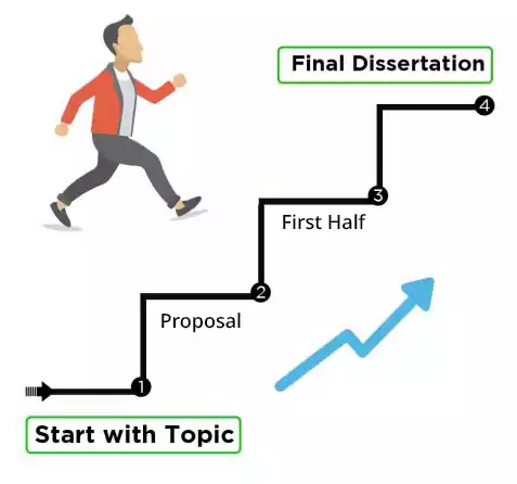
Final Words
You can use or get inspired by our selection of the best radiology diss. You can also check our list of critical care nursing dissertation topics and biology dissertation topics because these areas also relate to the discipline of medical sciences.
Choosing an impactful radiology dissertation topic is a daunting task. There is a lot of patience, time and effort that goes into the whole process. However, we have tried to simplify it for you by providing a list of amazing and unique radiology dissertation topics for you. We hope you find this blog helpful.
Also learn about our dissertation services here .
Free Dissertation Topic
Phone Number
Academic Level Select Academic Level Undergraduate Graduate PHD
Academic Subject
Area of Research
Frequently Asked Questions
How to find radiology dissertation topics.
For radiology dissertation topics:
- Research recent advancements.
- Identify unexplored areas.
- Consult experts and journals.
- Focus on patient care or tech.
- Consider ethical or practical issues.
- Select a topic resonating with your passion and career objectives.
You May Also Like
Disasters can potentially be quite dangerous to the continued existence of humans on Earth. Therefore, it is crucial to develop fresh, cutting-edge approaches to managing the damage caused by natural disasters.
Need interesting and manageable Instagram dissertation topics? Here are the trending Instagram dissertation titles so you can choose the most suitable one.
There are a wide range of topics in sports management that can be researched at the national and international levels. International sports are extremely popular worldwide, making sports management research issues very prominent as well.
USEFUL LINKS
LEARNING RESOURCES

COMPANY DETAILS

- How It Works
- Google Meet
- Mobile Dialer

Resent Search

Management Assignment Writing

Technical Assignment Writing

Finance Assignment Writing

Medical Nursing Writing

Resume Writing

Civil engineering writing

Mathematics and Statistics Projects

CV Writing Service

Essay Writing Service

Online Dissertation Help

Thesis Writing Help

RESEARCH PAPER WRITING SERVICE

Case Study Writing Service

Electrical Engineering Assignment Help

IT Assignment Help

Mechanical Engineering Assignment Help

Homework Writing Help

Science Assignment Writing

Arts Architecture Assignment Help

Chemical Engineering Assignment Help

Computer Network Assignment Help

Arts Assignment Help

Coursework Writing Help

Custom Paper Writing Services

Personal Statement Writing

Biotechnology Assignment Help

C Programming Assignment Help

MBA Assignment Help

English Essay Writing

MATLAB Assignment Help

Narrative Writing Help

Report Writing Help

Get Top Quality Assignment Assistance

Online Exam Help

Macroeconomics Homework Help

Change Management Assignment Help

Operation management Assignment Help

Strategy Assignment Help

Human Resource Management Assignment Help

Psychology Assignment Writing Help
Algebra Homework Help

Best Assignment Writing Tips

Statistics Homework Help

CDR Writing Services

TAFE Assignment Help

Auditing Assignment Help

Literature Essay Help

Online University Assignment Writing

Economics Assignment Help

Programming Language Assignment Help

Political Science Assignment Help

Marketing Assignment Help

Project Management Assignment Help

Geography Assignment Help

Do My Assignment For Me

Business Ethics Assignment Help

Pricing Strategy Assignment Help

The Best Taxation Assignment Help

Finance Planning Assignment Help

Solve My Accounting Paper Online

Market Analysis Assignment

4p Marketing Assignment Help

Corporate Strategy Assignment Help

Project Risk Management Assignment Help

Environmental Law Assignment Help

History Assignment Help

Geometry Assignment Help

Physics Assignment Help

Clinical Reasoning Cycle

Forex Assignment Help

Python Assignment Help

Behavioural Finance Assignment Help

PHP Assignment Help

Social Science Assignment Help

Capital Budgeting Assignment Help

Trigonometry Assignment Help

Java Programming Assignment Help

Corporate Finance Planning Help

Sports Science Assignment Help

Accounting For Financial Statements Assignment Help

Robotics Assignment Help

Cost Accounting Assignment Help

Business Accounting Assignment Help

Activity Based Accounting Assignment Help

Econometrics Assignment Help

Managerial Accounting Assignment Help

R Studio Assignment Help

Cookery Assignment Help

Solidworks assignment Help

UML Diagram Assignment Help

Data Flow Diagram Assignment Help

Employment Law Assignment Help

Calculus Assignment Help

Arithmetic Assignment Help

Write My Assignment

Business Intelligence Assignment Help

Database Assignment Help

Fluid Mechanics Assignment Help

Web Design Assignment Help

Student Assignment Help

Online CPM Homework Help

Chemistry Assignment Help

Biology Assignment Help

Corporate Governance Law Assignment Help

Auto CAD Assignment Help

Public Relations Assignment Help

Bioinformatics Assignment Help

Engineering Assignment Help

Computer Science Assignment Help

C++ Programming Assignment Help

Aerospace Engineering Assignment Help

Agroecology Assignment Help

Finance Assignment Help

Conflict Management Assignment Help

Paleontology Assignment Help

Commercial Law Assignment Help

Criminal Law Assignment Help

Anthropology Assignment Help

Biochemistry Assignment Help

Get the best cheap assignment Help

Online Pharmacology Course Help

Urgent Assignment Help

Paying For Assignment Help

HND Assignment Help

Legitimate Essay Writing Help

Best Online Proofreading Services

Need Help With Your Academic Assignment

Assignment Writing Help In Canada

Assignment Writing Help In UAE

Online Assignment Writing Help in the USA

Assignment Writing Help In Australia

Assignment Writing Help In the UK

Scholarship Essay Writing Help

University of Huddersfield Assignment Help

Ph.D. Assignment Writing Help

Law Assignment Writing Help

Website Design and Development Assignment Help
Radiology Thesis Research Topics
A dissertation, or thesis, is an integral part the Radiology curriculum. It can be called MD, DNB, or DMRD. For your convenience, we have tried to collect radiology thesis topics from different sources. Writing a Radiology thesis is not for everyone. There is no way around it so accept it and get on with it. #PhilosophyGyan!). Get started on your thesis as soon as you can. You can finish your thesis before the exams to avoid stress. Your thesis may need to be edited many times so be ready for this and plan your time accordingly.
Here are some tips for choosing the right topic and thesis in Radiology research:
- Prospective studies are more effective than retrospective ones.
- For your radiology thesis, choose a topic that is simple.
- You can choose a new topic if you're really interested in research and have a mentor to guide you. After you're done, make sure you publish your research.
- It is a good idea to stick with a topic for your thesis that won't take too much of your time in most cases.
- This does not mean you should abandon your thesis or 'Ctrl L + CtrlV' it from someone from another university. Writing your thesis is the first step in research methodology. Please do it honestly.
- However, don't spend too much time writing/collecting data to support your thesis.
- Don't put off preparing your thesis. Once you have been given a guideline, begin researching the topics and writing the review.
- Do not rush to finish your thesis until a few months before the exam.
- Some people have been unable to appear on the exam due to not having submitted their thesis on time. Do not take your thesis lightly.
- I will reiterate once more: Do not choose the thesis topic of someone else. Learn about the types of cases your Hospital treats. A good thesis on a common topic is better than one that is poorly written on a more obscure one.
List of Radiology Thesis Topics
- The state of the art in MRI for the diagnosis of hepatic focal lesion
- Multimodality imaging evaluation for sacroiliitis in patients newly diagnosed with spondyloarthropathy
- Multidetector computed Tomography in Oesophageal Varices
- The role of positron emission imaging tomography and computed tomography for the diagnosis of thyroid cancer
- Ultrasound elastography is used to evaluate focal breast lesions
- Assessment of traumatic spinal injuries: role of MRI diffusion tensor imagery
- Sonographic imaging for male infertility
- Comparative analysis of digital subtraction and color Doppler in patients with occlusive arterial diseases
- CT urography and haematuria: What is its role?
- Functional magnetic resonance imaging plays a vital role in brain tumor surgery safety
- Prediction of preeclampsia by Doppler uterine artery
- Evaluation of neonatal Cholestasis: Role of Doppler ultrasonography and gray scale
- Validity of MRI for diagnosis of congenital anorectal abnormalities
- Assessment of clubfoot: Role of sonography
- Diffusion MRI plays a role in the preoperative evaluation for brain neoplasms
- Pre-anaesthetic evaluation and laryngeal conditions.
- Study of intrauterine growth restriction: multivessel (arterial, venous) Doppler velocity
- Multiparametric 3tesla-MRI for suspected prostatic malignancy
- Sonography is an important tool for identifying benign nodules in the thyroid.
- Multiple sclerosis: Role of advanced magnetic resonance imaging sequences
- Evaluation of jaw lesions: role of multidetector computed Tomography
- Ultrasound and MR Imaging are important in the evaluation of Musculotendinous Pathologies of Shoulder Joint
- Perfusion computed tomography plays a role in the assessment of cerebral blood flow, blood volume, and vascular permeability for cerebral neoplasms
- MRI flow quantification is used to assess the most common csf flow abnormalities
- Diffusion-weighted MRI is important in the evaluation of prostate lesions. It also helps to determine histopathological correlation.
- CT enterography for evaluation of small bowel problems
- To detect recurrence, compare perfusion magnetic resonance imaging and magnetic resonance spectroscopy in post-radiotherapy treated gliomas.
- Evaluation of paediatric retroperitoneal masses using multidetector computed Tomography
- Multidetector computed tmography plays a role in neck lesions
- Indian population estimates standard liver volume
Topics for a Radiology dissertation
- Multislice CT scan, barium swallow and their role in the estimation of the length of oesophageal tumors
- Malignant Lesions-A Prospective Study.
- Ultrasonography is an important tool for the diagnosis of acute abdominal disease in children.
- Role of three dimensional multidetector CT hysterosalpingography in female factor infertility
- Comparative evaluation of multidetector computedtomography (MDCT), virtual tracheobronchoscopy, and fiberoptic traceo-bronchoscopy for airway diseases
- The role of multidetector CT for small bowel obstruction evaluation
- Sonographic evaluation of adhesive capsulitis in the shoulder
- Utility of MR Urography Versus Other Techniques in Obstructive Uropathy
- An MRI of the postoperative knee
- 64-slice multi detector computed tomography plays an important role in the diagnosis of mesenteric and bowel injury after blunt abdominal trauma.
- In the evaluation of focal liver lesion, sonoelastography is combined with triphasic computed Tomography
- Evaluation of the role of transperineal ultrasound and magnetic resonance imaging in urinary stress incontinence in women
- Multidetector computed morphographic features of abdominal hernias
- Ultrasound elastography is used to evaluate lesions in major salivary glands
- Female urinary incontinence: Transvaginal ultrasound and Magnetic Resonance Imaging
- Evaluation of colonic lesions using MDCT colonography and double contrast barium enema
- Role of MRI for diagnosis and staging urinary bladder carcinoma
- Children with febrile neutropenia: Spectrum of imaging findings
- Children with chest tuberculosis: Spectrum of radiographic appearances
- Computerized tomography plays a role in the evaluation of mediastinal masses during paediatrics
- Diagnosis of renal artery stenosis by comparison of multimodality imaging in diabetics
- Multidetector CT virtual Hysteroscopy is an important tool in the diagnosis of female infertility.
- Evaluation of Crohn's Disease: The role of multislice computed Tomography
- CT quantification of airway and parenchymal parameters using 64-slice MDCT in patients with chronic obstructive lung disease
- Comparative evaluation of MDCT versus 3t MRI in radiographically diagnosed jaw lesions.
- Evaluation of the diagnostic accuracy of ultrasonography, colour-Doppler sonography, and low dose computed Tomography in acute appendicitis
- Ultrasonography , magnetic resonance cholangio-pancreatography (MRCP) in assessment of pediatric biliary lesions
- Multidetector computed Tomography in Hepatobiliary Lesions
- Assessment of peripheral nerve lesions using high resolution ultrasonography (HRU) and colour Doppler
- Multidetector computed Tomography in Pancreatic Lesions
Thesis topics in DNB radiology
- Magnetic resonance perfusion weighted imagery & spectroscopy are used to grade gliomas by correlating the perfusion parameter of the lesion and the final histopathological grade
- Magnetic resonance assessment of abdominal tuberculosis.
- Low dose spiral HRCT for diffuse lung disease is useful in diagnosing
- Evaluation of endometrial lesion evaluations using dynamic contrast enhanced and diffusion-weighted magnetic resonance imaging
- Digital breast tomosynthesis and contrast enhanced digital mammography are both available for early diagnosis of breast lesions.
- Assessment of Portal Hypertension using Colour Doppler flow and magnetic resonance imaging
- Magnetic resonance imaging allows for the evaluation of musculoskeletal problems
- Diffusion magnetic resonance imaging is an important tool in the diagnosis of brain lesions that are neoplastic or inflammatory.
- Radiological spectrum of HIV-infected children with chest diseases High resolution ultrasonography for neck masses in children
- With surgical findings
- Evaluation of spinal trauma: Role of MRI
- Type 2 diabetes mellitus: Sonographic evaluation of the peripheral nerves
- Perfusion computed tomography plays a role in the evaluation neck masses and correlation
- Ultrasonography plays a role in diagnosing knee joint problems
- Ultrasonography plays a role in the evaluation of different causes of pelvic pain during the first trimester.
- The Evaluation of Diseases of the Aorta or its Branches: Magnetic Resonance Angiography's Role
- MDCT fistulography for evaluation of fistulas in Ano
- Multislice CT plays a role in the diagnosis of small intestinal tumors
- High resolution CT plays a role in the differentiation of benign and malignant pulmonary nodules among children
- Multidetector computed urography in the treatment of urinary tract disorders: A study
- High resolution sonography plays an important role in the assessment of the ulnar nerve for patients suffering from leprosy.
- Radiological pre-operative evaluation of malignant and locally aggressive musculoskeletal tumors using magnetic resonance imaging and computed tomography.
- In acute pelvic inflammatory diseases, the role of MRI and ultrasound
- In the evaluation of shoulder pain, ultrasonography is more effective than computed tomographicarthrography
- Multidetector Computed Tomography is an important tool for patients suffering from blunt abdominal trauma.
- Evaluation of breast lesions: The role of extended field-of-view sonography and compound imaging
- Multidetector CT, perfusion CT are used to evaluate focal pancreatic lesion.
- Assessment of breast masses using sono-mammography or colour Doppler imaging
- Evaluation of laryngeal masses: role of CT virtual laryngoscopy
- Triple phase multi-detector computed tomography in the liver masses
Radiology thesis topics for reference
- Ultrasound elastography is used to evaluate hepatic dysfunction in chronic liver disease.
- Assessment of hydrocephalus in children: Role of MRI
- Sonoelastography is an important tool in the diagnosis of breast lesions
- Patients with intracranial tumors: The impact of volumetric tumor doubling on survival
- Perfusion computed tomography plays a role in the diagnosis of colonic lesions
- Proton MRI spectroscopy plays a role in the evaluation and treatment of temporal lobe epilepsy
- Evaluation of peripheral arterial disease: role of multidetector CT and Doppler ultrasound
- Multidetector computed Tomography plays a role in paranasal sinus disease
- Virtual endoscopy with MDCT is an effective tool for diagnosing and evaluating gastric problems
- High resolution 3 Tesla MRI for the assessment of hindfoot and ankle pain.
- Ultrasonography transperineal in infants suffering from anorectal malformation
- In order to detect varices in patients with cirrhotics, CT portography uses MDCT instead of color Doppler
- CT urography plays a role in the evaluation of a dilapid ureter
- Dynamic contrast-enhanced multidetector CT characterizes pulmonary nodules
- Comprehensive CT imaging of an acute ischemic stroke using multidetector CT
- Fetal MRI plays a vital role in diagnosing intrauterine neurological congenital abnormalities
- Multidetector computed angiography plays a role in pediatric chest mass
- Multimodality imaging for the assessment of breast lesions that are palpable or non-palpable.
- Sonographic Assessment of Fetal Nasal Bone Length at 11-28 Gestational Days and Its Relationship to Fetal Outcome.
- The Role Of Sonoelastography and Contrast-Enhanced Computed Tomography in Evaluation Of Lymph Node Metastasis in Head and Neck Cancers
- Unenhanced computed Tomography allows for assessment of the hepatic fat in fatty liver disease.
- Correlation between vertebral marrow fat and spectroscopy, diffusion-weighted MRI imaging, and bone mineral density in postmenopausal females
- Comparative assessment of CT coronary imaging with conventional catheter coronary angiography
- Ultrasound evaluation of the length and diameter of the descending colon in normal and intrauterine-restricted foetuses
- Prospective study of the hepatic vein waveform in liver cirrhosis. Correlation with Child Pugh's classification.
- CT angiography for evaluation of coronary artery bypass graft patency in symptomatic patients' functional assessment myocardium using cardiac MRI in patients suffering from myocardial injury
- MRI Evaluation of HIV Positive Patients with Central Nerv System Manifestations
- MDCT evaluation of mediastinal hilar masses
- Evaluation of labro-ligamentous complex lesion by MRI & MRI arthrography shoulder joint
- Imaging plays a role in the assessment of soft tissue vascular malformations
Thesis topics in MD radiology:
- The Role of CT Virtual Cystoscopy in Urinary Bladder Neoplasia Diagnosis
- Multislice CT is an essential diagnostic technique for small intestinal tumours.
- "Mri Flow Quantification in the Evaluation of the Most Common CSF Flow Anomals"
- "The Fetal Mri Role in the Diagnosis of Intrauterine Neurological CongenitalAnomalies"
- Transcranial Ultrasound in the Diagnosis of Neonatal Brain Insults
- "Interventional Imaging Procedures' Role in the Treatment of Specific Gynecological Disorders"
- The Role of Radiological Imaging in Endometrial Carcinoma Diagnosis
- "The Role of High Resolution CT in the Diagnosis of Benign and Malignant Pulmonary Nodules in Children"
- Ultrasonography is a valuable diagnostic technique for knee joint pathologies.
- "The Role of Diagnostic Imaging Modalities in Assessing Post-Liver Transplantation Recipient Complications"
- "In Diagnosis, Diffusion-Weighted Magnet Resonance Imaging
- Brain Tumor Characterization in Relation to Conventional Mri
- PET-CT and Hepatic Tumor Evaluation
- "The Role of CT in the Evaluation of Mediastinal Masses in Pediatric Patients"
- "Female Urinary Incontinence: Transvaginal Ultrasound and Magnetic Resonance Imaging
- Multidetector CT is an important tool in diagnosing urinary bladder cancer
- "The Role Of Transvaginal Ultrasound in Diagnosis and Treatment Of Female Infertility
- Role Of Diffusion-Weighted Mri Imaging In Evaluation Of Cancer Prostate
- "Role Of Emission Tomography With Computed Tomography In Diagnosis Of Cancer Thyroid"
- CT Urography in the Case of Haematuria: What Role Does It Play?
- "The Role of Ultrasonography in the Diagnosis of Acute Abdominal Disorders in Children"
- "The Role of Functional Magnetic Resonance Imaging in Increasing the Safety of Brain Tumor Surgery"
- The Role of Sonoelastography in the Characterization of Breast Lesions
- "Ultrasonography and Magnetic Resonance Cholangiopancreatography (MRCP) in Pediatric Biliary Lesions"
- "The Role of Ultrasound and Color Doppler Imaging in the Evaluation of Acute Abdominal Pain Caused by Female Genital Causes"
- "The Role of Multidetector CT Virtual Laryngoscopy in the Diagnosis of Laryngeal Mass Lesions"
- The Postoperative Knee MRI
- Mri's Role in Valvular Heart Disease Assessment
- Fetal Abdominal Abnormalities: The Role of 3D and 4D Ultrasonography
- State-of-the-Art Hispatic Focal Lesions
Get Trustworthy Thesis Assistance Offerd From AHECounselling
Take a deep breath, and think about the issues. What are your problems when writing a thesis. Why can't your thesis be transferred to our thesis-helpers? Are you looking for additional assistance in writing your thesis? These concerns can be frustrating. However, losing your marks is not something you want. Consider all the reasons you should visit AHECounselling to get dissertation help.
We are proud to offer our thesis writing services and assist scholars with these problems. You will find qualified thesis helpers at AHECounselling that can meet all your needs. We can answer all your questions about any topic. Professional thesis assistance is also available to help with the drafting, editing, and proofreading. It is now time to click the Order Now button. Place your order quickly before it is too late to submit your thesis!
Frequently asked questions
How do i choose a thesis for my radiology .
Select a straightforward subject for your radiology thesis. You can pick a unique topic if you have a competent mentor who will help you and are really engaged in research. Once you've completed that, be sure to publish your study as soon as it's finished.
What are the problems in radiology ?
The "invisible" radiologist, tissue characterization, and micro resolution are among the problems. Opportunities exist in interventional radiology and quantitative imaging. Radiological screening practices will alter due to in vitro diagnostics. Radiology may have varied effects from automation.
What are the 5 most common errors in radiology ?
In 2016, Johnson found that failure to consult earlier studies or reports, limitations in imaging technique (inappropriate or incomplete protocols), inaccurate or incomplete history, the lesion's location outside of the region of interest, and a failure to search were the most frequent causes of diagnostic errors.
What do radiology means ?
Imaging technology is used in the medical specialty of radiology to identify and treat illness. Diagnostic radiology and interventional radiology are two subfields of radiology. Radiologists are medical professionals with a focus on radiology.
What is an example of radiology ?
The most typical kinds of radiological diagnostic tests include: The term "computed tomography" (CT) is also used for CAT scans, which include CT angiography. upper gastrointestinal and barium enema fluoroscopy. MRI and MR angiography are terms for magnetic resonance imaging.
Does radiologist do surgery ?
A surgical operation, for instance, may be supported by medical imaging used by an interventional radiologist. With the use of this imaging, operations may be performed more safely and with a quicker recovery. Typically, interventional radiologists do keyhole surgery.
What does the future hold for radiology ?
Future phases of AI in radiology will build sophisticated deep learning algorithms, more complicated artificial neural networks, and intricate integration of several data systems (pathology and radiology) so that AI in medicine and radiology will continue to advance and become more potent.
Is AI going to replace radiologists ?
Radiologists cannot be replaced by AI. However, it can make radiologists' routine work easier. Early adopters of AI will therefore probably lead the radiology industry in the future. Some radiology medical students have changed their perspectives in response to this topic, which has raised concerns.
Which field is better nursing or radiology ?
Radiologic technologists made an average yearly pay of $56,450 as of 2012, according to the BLS. This is significantly greater than the average yearly salary of LPNs and certified vocational nurses, which was $42,400. But the majority of nurses make more money than radiologic technologists.
How do radiology techs make more money ?
You will be paid extra if you select a shift that starts later in the day. You will get paid extra if you pick shifts on the weekends. A radiologic technician who works the night shift gets paid much more per hour than one who works the day shift.
Do radiologists talk to patients ?
Direct patient interaction is already a common practice in several radiology subspecialties. Before, during, and after tests, sonologists, fluoroscopists, interventional radiologists, women's imagers, and pediatric radiologists frequently speak with their patients directly.
How long does it take to become a radiologist ?
You must complete a minimum of seven years of formal medical education. A master's in radiology follows a bachelor's in radiography with a biology and physics emphasis, similar to an MBBS or premedical degree.
Can radiologist do pain management ?
Numerous operations that our radiologists may carry out can aid in the pain reduction of suffering individuals. Many of those procedures can be very beneficial for people with joint pain, back pain, or chronic face discomfort.
List of Radiology Thesis Topics ?
- The role of positron emission imaging tomography and computed tomography in the diagnosis of thyroid cancer
Radiology thesis topics for reference ?

Top 10 Best Universities Ranking list in India 2022

Generic Conventions: Assignment Help Services

Research Paper Topics For Medical

Top 5 Resources for Writing Excellent Academic Assignments

How to Write a Literature Review for Academic Purposes

Tips for Writing a killer introduction to your assignment

How To Write A Compelling Conclusion For Your University Assignment

Research Papers Topics For Social Science

Best 150 New Research Paper Ideas For Students

7 Best Plagiarism Checkers for Students And Teachers in 2024
Enquiry form.
- +44 7897 053596
- [email protected]

Get an experienced writer start working
Review our examples before placing an order, learn how to draft academic papers, radiography dissertation topics.

How to Write a Unique Dissertation Title: Guidance and Illustrations

How long should my dissertation be?

- Dissertation Topics

Radiography is the scientific technology of producing images of internal body organs and tissues. This revolutionary imaging technique of science has been widely used to diagnose issues of a body’s internal structure. Radiography is a helpful field for the medical diagnosis that requires extensive research. Students need to find exciting and up-to-date radiography dissertation topics .
Find Out Quality Biomedical Science Dissertation Examples
Premier Dissertations has produced a list of new dissertation topics in radiography for 2024 .
If you would like to choose any topic from the list below, simply drop us a WhatsApp or an Email .
You may also like to review;
Healthcare Management Dissertation Topics | Pharmacy Dissertation Topics
How Does It Work ?

Fill the Form
Please fill the free topic form and share your requirements

Writer Starts Working
The writer starts to find a topic for you (based on your requirements)


3+ Topics Emailed!
The writer shared custom topics with you within 24 hours
List of Latest Radiography Research Topics 2024
Top thesis topics in radiography topics 2024, trending research topics in radiography dissertation topics, a methodical approach to choose a good radiography dissertation topic.
Selecting radiology research topics involves a methodical approach. Start by identifying your specific interests within radiography, such as diagnostic imaging, radiation therapy, or advancements in technology. Formulate a clear research aim and methodology, ensuring a focused and insightful exploration of your chosen area to contribute meaningfully to the field of radiography.
Review Our Full List of Latest Research Topics
For more radiography thesis topics and radiology research paper topics , please keep checking our website as we keep adding new topics to our existing list of titles. GOOD LUCK!
Get 3+ Free Radiography Dissertation Topics within 24 hours?
Your Number
Academic Level Select Academic Level Undergraduate Masters PhD
Area of Research
Get an Immediate Response
Discuss your requirments with our writers
Discover More:
Testimonials
Very satisfied students
This is our reason for working. We want to make all students happy, every day. Review us on Sitejabber
admin farhan
Related posts.

Controversial Psychology Topics

70 Visual Aid Speech Topics for Your Next Presentation

100+ Quantitative Research Titles and Topics
Comments are closed.
Home » Blog » Dissertation » Topics » Radiology » 80 Radiology Research Topics

80 Radiology Research Topics
FacebookXEmailWhatsAppRedditPinterestLinkedInIf you are a researcher in the field of Radiology searching for intriguing research topics, you’ve landed on the right platform. Selecting the proper research topics is pivotal to the success of their thesis or dissertation. From exploring cutting-edge technologies to enhancing diagnostic accuracy, radiology constantly evolves, presenting students with an exciting spectrum of potential […]

If you are a researcher in the field of Radiology searching for intriguing research topics, you’ve landed on the right platform. Selecting the proper research topics is pivotal to the success of their thesis or dissertation. From exploring cutting-edge technologies to enhancing diagnostic accuracy, radiology constantly evolves, presenting students with an exciting spectrum of potential research topics for their undergraduate, master’s, or doctoral-level theses and dissertations.
Radiology, also known as “radiography,” “medical imaging,” and “diagnostic imaging,” often referred to as medical imaging, is a specialized branch of medicine concerned with utilizing various imaging techniques to diagnose and treat medical conditions.
A List Of Potential Research Topics In Radiology:
- Evaluating the impact of radiological research on public health policies and healthcare decision-making.
- Assessing the diagnostic accuracy of diffusion tensor imaging (DTI) in evaluating spinal cord injury.
- Evaluating the role of 18F-FDG PET/CT in assessing treatment response in soft tissue sarcomas.
- Investigating the role of radiogenomics in predicting genetic biomarkers associated with glioblastoma multiforme.
- Analyzing the contributions of radiology in early disease detection and prevention strategies.
- Investigating the potential of radiogenomics in predicting response to immunotherapy in lung cancer patients.
- Investigating the potential of radiogenomics in predicting response to targeted therapies in breast cancer.
- Evaluating the effectiveness of artificial intelligence algorithms in detecting and characterizing thyroid nodules on ultrasound.
- Evaluating the utility of perfusion MRI in assessing treatment response in patients with intracranial tumors.
- Assessing the diagnostic performance of PET/MRI in evaluating brain tumors and treatment planning.
- Analyzing the impact of multiparametric MRI in the early detection and staging of aggressive prostate cancer.
- Evaluating the role of virtual colonoscopy (CT colonography) in colorectal cancer screening and surveillance.
- Evaluating the efficacy of telemedicine in delivering radiology consultations and follow-up care in a post-COVID healthcare landscape.
- Exploring the interplay between radiology and psychology in diagnostic decision-making.
- Investigating the potential of radiomics in predicting survival outcomes and treatment response in glioblastoma patients.
- Evaluating the role of advanced imaging techniques in assessing treatment response in patients with pancreatic neuroendocrine tumors.
- Assessing the socioeconomic factors influencing the accessibility and utilization of radiological services worldwide.
- The ethical considerations in radiology research include patient consent, radiation exposure, and data privacy.
- Assessing the environmental sustainability of radiology practices in the UK, including reducing radiation exposure and waste.
- Assessing the diagnostic performance of positron emission tomography/magnetic resonance imaging (PET/MRI) in prostate cancer.
- Analyzing the impact of diffusion-weighted imaging (DWI) in assessing treatment response in rectal cancer patients.
- Investigating the role of radiology in precision medicine and personalized treatment plans.
- Understanding the implications of virtual reality-based training for improving interventional radiology skills and performance.
- Investigating the potential of radiomics in predicting treatment response and survival outcomes in hepatocellular carcinoma.
- Assessing the diagnostic performance of dual-energy CT in evaluating gout and other crystal arthropathies.
- Understanding the impact of low-dose CT protocols on lung cancer screening and radiation exposure.
- Analyzing the role of perfusion imaging in assessing treatment response in patients with brain metastases.
- Assessing the diagnostic accuracy of dual-energy CT in characterizing renal masses and guiding treatment decisions.
- Assessing the accessibility and equity of radiological services in different regions of the UK.
- Investigating the role of radiogenomics in predicting response to immunotherapy in patients with melanoma.
- Assessing the role of artificial intelligence in optimizing radiological image interpretation and diagnosis post-COVID-19.
- Studying the challenges and opportunities of applying radiology in global health initiatives and disaster response.
- Investigating the role of radiology in diagnosing and managing common health issues in the UK, such as cardiovascular diseases.
- Analyzing the long-term consequences of delayed or deferred radiological procedures during the COVID-19 crisis.
- Evaluating the effectiveness of 3D printing in surgical planning for complex craniofacial reconstructions.
- Evaluating the diagnostic accuracy of dual-energy CT in identifying renal stone composition and guiding treatment strategies.
- Evaluating the effectiveness of artificial intelligence algorithms in detecting and characterizing pulmonary nodules on chest CT.
- Investigating the integration of artificial intelligence and machine learning in the National Health Service (NHS) radiology services.
- Evaluating the role of diffusion tensor imaging (DTI) in assessing white matter changes in patients with Alzheimer’s disease.
- Exploring the ethical and legal implications of data sharing and privacy concerns in radiology research related to COVID-19.
- Analyzing the development of radiology as a medical specialty and its interdisciplinary collaborations over the years.
- Analyzing the impact of diffusion-weighted MRI in characterizing ovarian tumors and guiding treatment decisions.
- Investigating the adoption of advanced imaging techniques for monitoring post-COVID-19 complications in recovering patients.
- Exploring the use of radiology in managing healthcare disparities among different socioeconomic groups in the UK.
- Analyzing the potential of spectral CT imaging in differentiating benign and malignant thyroid nodules.
- Investigating the impact of the COVID-19 pandemic on radiology department workflows and patient management.
- Reviewing the historical evolution of radiological imaging techniques and their contributions to medical diagnostics.
- Analyzing the effectiveness of artificial intelligence algorithms in automated segmentation of liver lesions on CT scans.
- Assessing the diagnostic accuracy of dual-energy CT in differentiating benign and malignant adrenal lesions.
- Investigating the potential of radiogenomics in predicting treatment response in glioma patients undergoing radiotherapy.
- Evaluating the effectiveness of quantitative MRI in characterizing cartilage changes in osteoarthritis patients.
- Analyzing the impact of artificial intelligence algorithms in automated segmentation of lung lesions on CT scans.
- Assessing the diagnostic accuracy of 68Ga-PSMA PET/CT in detecting and localizing recurrent prostate cancer.
- Assessing the diagnostic accuracy of magnetic resonance elastography (MRE) in liver fibrosis staging.
- Assessing the diagnostic performance of multimodal imaging in the evaluation of musculoskeletal infections.
- Evaluating the role of artificial intelligence in automated classification of breast lesions on mammography and ultrasound.
- Exploring radiologists’ psychological well-being and stress levels after the COVID-19 pandemic.
- Analyzing the impact of deep learning algorithms in the automatic segmentation of brain tumors on MRI.
- Assessing the utilization of radiology in tracking the progression of post-acute sequelae of SARS-CoV-2 infection (PASC).
- Investigating the potential of radiomics in predicting response to neoadjuvant chemotherapy in esophageal cancer.
- Analyzing the role of artificial intelligence in improving the accuracy and efficiency of mammography interpretation.
- Investigating the potential of radiogenomics in predicting response to neoadjuvant chemotherapy in breast cancer.
- Evaluating the effectiveness of radiological interventions in managing infectious disease outbreaks, focusing on lessons from the UK’s response to epidemics.
- The impact of health policy on radiology: a comprehensive analysis of regulations and access to care.
- Analyzing the role of radiology in supporting the aging population and addressing age-related health challenges in the UK.
- Analyzing the impact of Brexit on radiology research collaborations and data sharing with European institutions.
- Studying the impact of COVID-19 vaccination campaigns on radiological findings in the UK, including vaccine-related complications.
- Investigating the role of radiomics in predicting treatment response and survival outcomes in pancreatic cancer.
- Analyzing the effectiveness of image-guided ablation therapies for liver tumors in improving patient outcomes.
- Studying the use of radiological imaging in diagnosing and managing multisystem inflammatory syndrome in children (MIS-C) post-COVID-19.
- Analyzing the potential of radiomic features in predicting lymph node metastasis in breast cancer patients.
- Assessing the diagnostic accuracy of MRI-guided breast biopsy in patients with suspicious lesions detected on mammography.
- Investigating the potential of 4D flow MRI in assessing cardiovascular hemodynamics and guiding surgical interventions.
- Evaluating the effectiveness of national health policies and initiatives in promoting early cancer detection through radiological screening in the UK.
- Analyzing the economic impact of COVID-19 on radiology departments and the implementation of cost-effective strategies.
- Understanding the implications of contrast-enhanced ultrasound in diagnosing and monitoring liver fibrosis progression.
- Investigating the impact of advanced imaging techniques on early detection and treatment outcomes of breast cancer.
- Assessing the effectiveness of radiographic measures in monitoring disease progression and treatment response in rheumatoid arthritis.
- Evaluating the effectiveness of artificial intelligence algorithms in diagnosing acute stroke using CT and MRI.
- Analyzing the impact of quantitative imaging biomarkers in predicting treatment response in esophageal cancer patients.
In conclusion, choosing the right research topic in radiology for your thesis or dissertation is an opportunity to delve into the dynamic and ever-evolving field of medical imaging. The selected research topic should align with your academic level and interests, whether exploring the potential of artificial intelligence in radiology, optimizing imaging techniques, or investigating the latest advancements in the field. Remember, your research journey should be both fulfilling and contribute meaningfully to the world of radiology.
Order Radiology Dissertation Now!
External Links:
- Download Radiology Dissertation Sample For Your Perusal
Research Topic Help Service
Get unique research topics exactly as per your requirements. We will send you a mini proposal on the chosen topic which includes;
- Research Statement
- Research Questions
- Key Literature Highlights
- Proposed Methodology
- View a Sample of Service
Ensure Your Good Grades With Our Writing Help
- Talk to the assigned writer before payment
- Get topic if you don't have one
- Multiple draft submissions to have supervisor's feedback
- Free revisions
- Complete privacy
- Plagiarism Free work
- Guaranteed 2:1 (With help of your supervisor's feedback)
- 2 Installments plan
- Special discounts
Other Posts
WhatsApp us
Selected Project topics in Medical Radiography And Radiological Sciences
We have randomly selected Current Project topics in Medical Radiography and Radiographical Science fields.
These topics can be developed in many ways and we have materials for these topics listed and more to guide you throughout your research work.
These topics include:
1. Pattern Of Findings In The Echocardiogram Of Hypertensive Patients In Conquest Medical Imaging Centre Enugu Background: Echocardiography has become and important tool in medical imaging. it is used in the confirmation of heart diseases especially hypertension, in nigeria major cities using conventio… Premium 79 pages 12936 words Project
2. Evaluation Of Compliance With Standard Postero–anterior (pa) Chest Techniques This study was carried out to evaluate the compliance with standard postero-anterior (PA) chest Techniques in University of Nigeria Teaching Hospital Ituku Ozalla. A total of 375 assessm… Premium 83 pages 11628 words Project
3. Identifying The Factors Influencing The Attitude Of Radiographers To Their Professional Body A professional association is usually a non-profit organization seeking to further a particular profession, the interests of individuals engaged in that profession and the public. The roles of… Premium 61 pages 10799 words Project
4. Local Construction Of Battery Powered Dual Face Radiographic Viewing Box With Rotatable Neck And Adjustable Stand The research work was designed to construct a locally battery powered dual face radiographic viewing box with rotatable neck and adjustable stand. The illumina… Premium 78 pages 12131 words Project
5. Public Awareness Of The Health Effect Of Radiation Emitted From Telecommunication Mast This study proposes to research the awareness of the public on the health effect of radiation emitted from telecommunication mast. Since the major effect of this radiation emitted from telecommunicati… Premium 72 pages 11350 words Project
6. Royal College Of Radiologists Guidelines For Abdominal Radiography Requests: Assessment Of Referring Clinicians’ Adherernce Plain abdominal radiography is believed to be over utilized as an investigation for acute abdominal complaints. Because of this, the Royal College of Radiologists published guidelines in order… Premium 38 pages 4520 words Project
7. Establishment And Management Of A Private Ultrasonographic Centre In Enugu Metropolis; Problems And Prospects ABSTRACT This study was done to determine the problems and prospects of setting up and running a private ultrasonographic centre in Enugu metropolis. It was carried out in the 38 registered diagnostic … Premium 64 pages 9530 words Project
8. Correlation Of Mri Findings With Outcome Of Treatment In Patients With Spinal Cord Injury: A Case Study Of Lagos University Teaching Hospital Idi-araba Lagos Nigeria ABSTRACT Study DesignA retrospective studyObjectivesTo correlate the MRI findings with the outcome of patient treatment in patients with spinal cord injury. This study tried to study the recovery of pa… Premium 66 pages 11037 words Project
9. Common Magnetic Resonance Imaging Findings In Patients With Neurologic Disorders, In University Of Ilorin Teaching Hospital, Kwara State ABSTRACT Objective: To evaluate the common MRI findings in patients with neurologic disorders.Method: A retrospective study of 106 patients with neurologic disorders was carried out and their respectiv… Premium 99 pages 16464 words Project
10. Characterization Of Breast Lesions Seen During Mammography At University Of Nigeria Teaching Hospital Enugu ABSTRACT This research was carried out to characterize the variety of the breast lesion seen during mammography at UNTH and to establish the age distribution of the lesion at the time of presentat… Premium 49 pages 8508 words Project
11. Prevalence Of Low Back Pain Among Practicing Radiographers In Enugu And Ebonyi States (a Case Study Of Unth, Nohe, Esuth And Fetha) ABSTRACT This study on the prevalence of low back pain among practicing radiographers in government hospitals, was done in Enugu and Ebonyi States. The objectives of the study were to determine t… Premium 57 pages 9304 words Project
12. Establishment And Management Of A Private Radio-diagnostic Centre In Onitsha, Anambra State Problems And Prospects. ABSTRACT This study was done to determine the problems and prospects of establishing and managing a private radio-diagnostic centre in Onitsha, Anambra state. It was carried out in the 42 diagnostic c… Premium 67 pages 9276 words Project
13. Assessment Of The Perspective And Attitude Of Radiology Staff To Radiography Students For Clinical Training In The Radiology Department (a Case Study Of Unth, Esuth And Nohe) ABSTRACT Clinical training of radiography students is part of the medical academic curriculum in a hands-on environment where students are taught skills, behaviors and attitudes required to enter into … Premium 70 pages 13620 words Project
14. Assessment Of Mammographic Screening Awareness Among Female Traders In Enugu Metropolis, Enugu State ABSTRACT So far, mammographic screening is the only mode of detection that has been shown to reduce breast cancer mortality (Montazeri et al). Thus unless women are educated about mammography and… Premium 61 pages 12414 words Project
FOR MORE SELECTED TOPICS IN THIS FIELD, KINDLY CLICK HERE
Share this:
9 thoughts on “ selected project topics in medical radiography and radiological sciences ”.
please I need. more light on selecting project topic on xray department and preparable topic to be selected
please I need more researchable topics on mammography
Pleace o need a researchable topics on compited tomography
Please go to https://afribary.com/works?search=Computed+Tomography
Please i need aresearch topic in in the area of xray
Please need a researchable topic in ultrasound examination and the machine itself
You really done a wonderful job. Thank you and God bless
Pls need a research on medical imaging technology project work for student
Please I need project topic on radiographic cassette or film
Leave a Comment Cancel reply
Your email address will not be published. Required fields are marked *
Notify me of follow-up comments by email.
Notify me of new posts by email.
This site uses Akismet to reduce spam. Learn how your comment data is processed .
- Brownell Lab
- Brugarolas Lab
- Elmaleh Lab
- Johnson Lab
- Normandin Lab
- Santarnecchi Lab
- Sepulcre Lab
- Technical Staff
- Administrative Staff
- Collaborators
- Publications
Image Generation and Clinical Assessment
- PET and SPECT Instrumentation
Multimodal MRI Research
Quantitative pet-mr imaging.
- Quantitative PET/SPECT
Machine Learning for Anatomical imaging
- Therapy Imaging Program
Pharmaco-Kinetic Modeling
Monitoring radiotherapy with pet.
- Radiochemistry Discovery
- Precision Neuroscience & Neuromodulation Program
- Video Lectures
- T32 Postgraduate Training Program in Medical Imaging (PTPMI)
- The Gordon Speaker Series
- Translation
- Covid-19 response
- Recent Seminars
- Awards and Honors
- Recent Publications
- Other News Items
- Grant Proposals
- Grant Related FAQs
- Travel Policies
- Purchasing and Other Daily Operations
- Human Resources
- Contact Forms
- Science Resources
- Computing Resources
- File Repository
- Seminar Sign Up

Research Topics
The Gordon Center is conducting work at the forefront of quantitative PET in brain, oncologic and cardiac imaging. This webpage summarizes some of the research performed in the Center in kinetic modeling of neurotransmission, cardiac perfusion and mitochondrial function, simultaneous PET-MR imaging, in-room PET monitoring of proton therapy, high sensitivity/resolution brain imaging, quantitative dual tracer PET and SPECT, and objective assessment of image quality for estimation and detection tasks. More information about our research areas can be found in the links below.
PET-CT and SPECT Instrumentation
Quantitative PET-CT and SPECT-CT
Therapy Imaging Program (TIP)
Radiochemistry Discovery and Multifunctional Nanomaterials
Precision Neuroscience & Neuromodulation Program
Concepts for exploring research avenues in radiology: opportunities and inspiration
- Published: 15 April 2023
- Volume 33 , pages 6545–6547, ( 2023 )
Cite this article
- Teodoro Martín-Noguerol 1 ,
- Suyash Mohan 2 &
- Antonio Luna 1
1608 Accesses
1 Altmetric
Explore all metrics
Avoid common mistakes on your manuscript.
Performing scientific research and publishing the results in peer-reviewed journals is vital to the advancement of medicine. In medical imaging, scientific research should be an integral part of the continuing education of radiologists. Research in radiology (or any other field of medicine) represents an international currency that transcends political borders, helps radiologists globally keep up to date with current advances, complements teaching, improves resident training, and enhances patient care [ 1 ].
Drawing up a research project is, in most cases, the result of a deep understanding of the purpose and expected outcomes related to a specific topic. However, it is not uncommon to become discouraged when looking at a blank page (or computer screen) or contemplating if the work is novel, whether it is relevant, or if it provides new information. This lack of inspiration may cause many radiologists to relinquish their research efforts.
Needless to say, radiologists need to be constantly submerged and actively involved in continuous educational and ongoing research activities in this ever-changing landscape of medicine to accomplish two main goals: address the issues that radiologists have to face in their daily clinical and radiological practice and stay up-to-date in all the innovations that are continuously emerging not only related to radiology but also in other medical or even social disciplines. Search engines such as PubMed or Google Scholar may help scientific communities in several ways from research to keeping bibliographies up to date. Social networks can also help radiologists stay informed about current trends by means of following general or specific radiologic society accounts or social media profiles. Along the same lines, virtual radiology meetings and webinars have experienced considerable growth in the last decade and are almost an endless supplier of resources not only for teaching and learning but also for developing ideas about research topics in radiology and its subspecialties. These scenarios can serve as the optimal breeding ground as a starting point to conceive promising research projects and address unresolved questions.
A deep understanding of what questions have been already answered and what issues still need to be fully explained or have not been adequately covered will for sure assist in the selection of relevant topics for further research, especially for younger radiologists under guidance from more senior experts.
Personal and institutional expertise can also help in identifying potential research projects in radiology, playing mentorship a crucial role to guide less experienced radiologists [ 2 ]. Radiology department seminars and hospital interdisciplinary conferences may also open a door to potential synergistic collaborations with other specialists, discovering specific needs not previously covered, and thus, finding innovative ideas and opportunities for scientific pursuit. In other words, thinking outside the “radiology box” and focusing on what other specialties need for improving their clinical practice and their patients’ outcomes and how can radiology play a role in answering these questions help in carving ideas for meaningful and translational research. The institutional work environment and available infrastructure is also an important consideration when identifying potential topics and ideas for research. Hospitals with a high prevalence of cases of a specific disease or tertiary care-referral hospitals can provide much-needed information to institutions and hospitals with limited experience in those specific clinical areas and thus enhancing patient care locally, and simultaneously providing radiologists a potential source for original scientific research projects. Along the same lines, if your institution or radiology department is a referral center for specific or advanced diagnostic or interventional techniques (already implemented or even under development), it can also be explored and exploited to obtain ideas for research and dissemination of knowledge to other centers.
In some cases, the source of inspiration or opportunities may even be found outside the work environment. Visiting other radiology departments or away rotations can open avenues and provide insights about how other colleagues address a specific task and how these can be imported into their home institutions. Of course, these kinds of experiences may enhance cooperative working between institutions and partners and thus, boost the possibilities of promoting collaborative science in radiology [ 3 ]. In some cases, national, international, or even regional radiology societies launch outreach programs and projects to address specific needs as identified by their expert committees in which radiologists may participate as researchers, as global challenges (e.g., COVID-19) require global solutions.
Scientific journals may also launch a call for papers asking to submit proposals for consideration for research and publication in a special issue dedicated to a specific topic. These proposals are usually endorsed into a focused issue and cover a spectrum of related topics across multiple radiology subspecialties, including areas such as practice management, quality, and safety. This scenario provides a potential opportunity for authors and can assist with channeling or tailoring their research ideas to fit into the editor’s requirements.
The global and scientific community, and by extension, the world, is in a state of continuous change and update. There are new technologies or trends in radiology , such as all topics related to artificial intelligence (AI), including the recent hype related to ChatGPT, and new emerging diseases that need radiology and imaging such as the COVID-19, where research interest has grown exponentially in the last few years [ 4 , 5 ]. AI and COVID-19 are just some examples of new niches that were previously unknown and that can be explored and exploited as potential sources of ideas for research in radiology [ 6 ]. At this point, a critical view of what radiologists do and what can they do is essential to find answers and shed light on important research topics in a meaningful way to eventually improve outcomes, healthcare, and radiology practice in general. For example, trying to export radiologic techniques from one anatomic region to another is also an interesting topic currently used to generate innovative ideas for research in radiology. For example, well-tested and proven techniques such as diffusion-weighted imaging, which was primarily developed for central nervous system imaging, have been successfully exported to other anatomic sites and organs such as the liver, prostate, or breast [ 7 ]. Nowadays there are still dozens of advanced radiologic technologies waiting to be explored and tested with applications different for which they were initially created and can help in providing answers or filling gaps in knowledge to previously scarcely addressed imaging challenges, clinical issues, or patients’ needs.
Nevertheless, ideas for research in radiology should be broad and not just limited to the clinical or purely radiological-image-based scope. As mentioned above, we are experiencing continuous changes in society with a real and worldwide impact. For this reason, our research should also be focused outside our comfort zone with the aim to explore and provide newer insights and potential solutions by tackling other usually neglected topics that could be related to economics (i.e., climatic change and reducing energy-related costs for radiology equipment), health equity, inclusion and diversity (i.e., gender differences or global access to screening programs), or resources management (i.e., a worldwide shortage of radiologists or facing with the “feared” integration of AI) among others.
Finally, the possibility of successfully identifying ideas for research in radiology depends on a strong team you can count on, which includes experts, PhD scientists, physicists, and coordinator support staff. Steady support and a collaborative working environment, mentorship, collegiality with other radiologists from the same department or different institutions, other clinical specialists, radiographers, engineers, data analysts, or the support of scientific societies or stakeholders will facilitate the development of new ideas with greater chances of success [ 8 ].
In conclusion, ideas and opportunities for conducting research in radiology are plentiful. They usually arise from the radiologist’s individual professional experience and their observations of the world around them. Inspiration will only emerge in a fertile environment of continuous research and study, or in the words of Pablo Picasso (Málaga 1881, Mougins 1973), one of the greatest avant-garde artists of all time, “inspiration exists, but it has to find you working.”
Bluemke DA (2018) The power of publishing. Radiology 286:735–736
Article Google Scholar
Awan OA, Katz DS (2021) Mentorship in radiology and in life. Radiographics 41:E98–E99
Article PubMed Google Scholar
Attyé A (2019) Data sharing improves scientific publication: example of the “hydrops initiative.” Eur Radiol 29:1959–1960
Bluemke DA (2018) Editor’s note: Publication of AI research in radiology. Radiology 289:579–580. https://doi.org/10.1148/radiol.2018184021
Biswas S (2023) ChatGPT and the future of medical writing. Radiology 223312. https://doi.org/10.1148/radiol.223312
Moy L, Bluemke DA (2021) The COVID-19 pandemic and radiology submissions. Radiology 298:483–484
Luna A, (Antonio), Ribes R, Soto JA (2012) Diffusion MRI outside the brain : a case-based review and clinical applications. Springer-Verlag, Berlin Heidelberg
Book Google Scholar
Martín-Noguerol T, Paulano-Godino F, López-Ortega R et al (2021) Artificial intelligence in radiology: relevance of collaborative work between radiologists and engineers for building a multidisciplinary team. Clin Radiol 76:317–324. https://doi.org/10.1016/j.crad.2020.11.113
Download references
The authors state that this work has not received any funding.
Author information
Authors and affiliations.
MRI Unit, Radiology Department, HT Medica, Carmelo Torres 2, 23007, Jaén, Spain
Teodoro Martín-Noguerol & Antonio Luna
Division of Neuroradiology, Hospital of the University of Pennsylvania, Philadelphia, PA, USA
Suyash Mohan
You can also search for this author in PubMed Google Scholar
Corresponding author
Correspondence to Teodoro Martín-Noguerol .
Ethics declarations
The scientific guarantor of this publication is Dr. Antonio Luna.
Conflict of interest
The authors of this manuscript declare relationships with the following companies: Antonio Luna, MD, PhD, is an occasional lecturer of Philips, Siemens Healthineers, Bracco, and Canon and receives royalties as a book editor from Springer-Verlag.
The rest of the authors of this manuscript declare no relationships with any companies, whose products or services may be related to the subject matter of the article.
Statistics and biometry
No complex statistical methods were necessary for this paper.
Informed consent
Written informed consent was not required for this study because the manuscript is an Editorial.
Ethical approval
Institutional Review Board approval was not required because the manuscript is an Editorial.
Study subjects or cohorts overlap
Methodology, additional information, publisher's note.
Springer Nature remains neutral with regard to jurisdictional claims in published maps and institutional affiliations.
Rights and permissions
Reprints and permissions
About this article
Martín-Noguerol, T., Mohan, S. & Luna, A. Concepts for exploring research avenues in radiology: opportunities and inspiration. Eur Radiol 33 , 6545–6547 (2023). https://doi.org/10.1007/s00330-023-09639-4
Download citation
Received : 20 February 2023
Revised : 20 February 2023
Accepted : 15 March 2023
Published : 15 April 2023
Issue Date : September 2023
DOI : https://doi.org/10.1007/s00330-023-09639-4
Share this article
Anyone you share the following link with will be able to read this content:
Sorry, a shareable link is not currently available for this article.
Provided by the Springer Nature SharedIt content-sharing initiative
- Find a journal
- Publish with us
- Track your research
404 Not found
Unveiling Trends in Radiology Science and Education | RSNA 2023
Find rsna 2023 sessions highlighting the latest advances in medical imaging.

RSNA 2023: Leading Through Change will bring together an international community of medical imaging professionals and industry partners and will help you stay up to date with the latest radiology advancements.
Featuring over 300 educational courses and more than 3,500 scientific papers, education exhibits and science posters, RSNA 2023 will also include an engaging line-up of plenary speakers sharing insights on the topics that are most relevant to today’s radiologists.
This year, the Technical Exhibits halls are home to more than 650 exhibitors, including over 100 first-time exhibitors. This group of welcome newcomers is comprised of recruiters and academic institutions as well as a variety of industry partners who are excited to share their products and services with you. Come see their offering of next-generation imaging modalities, AI, interoperability, workflow, 3D printing, automation and staffing solutions.
To help attendees plan for RSNA 2023, Jorge Soto, MD, chair of the RSNA Annual Meeting Program Planning Committee (AMPPC) provides a preview of this year’s trending topics. Dr. Soto notes that regardless of your subspecialty or career stage, there is something for everyone at RSNA 2023.
“Members of our committee performed an exhaustive review of the hundreds of abstract submissions,” Dr. Soto said. “It is exciting to have a first-hand look at the breadth and depth of work being done by our global colleagues and to realize the impact this work might have on our profession.”
Watch Dr. Soto discuss this year's RSNA 2023 meeting highlights:

In addition to a strong response to the initial call for abstracts, many submissions were received for a second abstract call that focused on late breaking research in the areas of generative AI, sustainability in imaging, imaging of immunotherapy and theranostics.
Sessions selected from the second call will be featured in the Learning Center theaters throughout the week. “After introducing the Learning Center Theater last year for the presentation of research selected during our second call, we are pleased to announce the addition of a second Learning Center Theater to accommodate schedules and seating for this popular new feature,” Dr. Soto said.
Which topics are trending for RSNA 2023?
“Trends vary somewhat, depending on your subspecialty, but applications of AI and photon-counting CT have been popular across nearly every subspecialty,” Dr. Soto said. “Theranostics is also increasing in popularity along with the use of large language models for a variety of different uses.”
Attendees will also enjoy a wide selection of non-interpretive sessions that will help them sharpen skills beyond imaging. With radiology professionals continuing to focus on ensuring equitable access to health care for all patient populations and understanding diversity, equity and inclusion, sessions throughout the week will be available for attendees interested in making changes at their own institutions and practices.
“We’ll have several experts sharing useful insights and ideas for improving in the areas of DEI and health equity,” Dr. Soto said. “These sessions can be thought-provoking both from the research and clinical perspectives.”
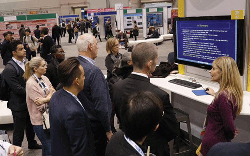
Dr. Soto noted that in addition to the latest research, attendees can also rely on the annual meeting for a robust offering of CME opportunities. “Both in-person and virtual participants will experience an immersive week filled with chances to discover, connect, learn and grow,” Dr. Soto said. “We look forward to providing this world-class experience to our world-class attendees.”
Wondering where to get started? Use this handy quick-reference guide developed with the assistance of AMPPC members to find trending topics and recommended sessions by subspecialty.
Trends by Subspecialty Practice Area
Breast Imaging
Cardiac Imaging
Chest Imaging
Gastrointestinal Imaging
Head & Neck Imaging
Imaging Informatics
Interventional Radiology
Multisystem Imaging
Musculoskeletal radiology.
Noninterpretive/Practice Management
Nuclear Medicine and Molecular Imaging
Neuroradiology
Pediatric Radiology
Radiation Oncology
OB/Gynecology Imaging
Vascular Imaging
Associated Sciences/ASRT
Look for these additional topics captivating attention in radiology .
For More Information
Register for the meeting.
Review the RSNA 2023 Program .
Read RSNA News stories about RSNA 2023:
- RSNA 2023 Technical Exhibits Highlight Latest Medical Imaging Innovations
- Registration Is Open for RSNA Labs
Annual Meeting Planning Committee
Information for this preview was provided by annual meeting program planning committee members:
Jorge A. Soto, MD, chair
Stamatia V. Destounis, MD, chair
Hiroyuki Abe, MD
Wendy B. Demartini, MD
Thomas H. Helbich, MD, MBA
Cherie M. Kuzmiak, DO
Katja Pinker-Domenig, MD, PhD
Karen G. Ordovas, MD, MS, chair
Carole J. Dennie, MD, FRCPC
Diana Litmanovich, MD
Michael F. Morris, MD
Ming-Yen Ng, MBBS
Prabhakar Rajiah, MD, FRCR
Ioannis Vlahos, FRCR, MBBS,
Saurabh Agarwal, MD
Kristopher W. Cummings, MD
Travis S. Henry MD
Jane P. Ko, MD
Anastasia Oikonomou, MD, PhD
Emergency Radiology Imaging
Manickam Kumaravel, MD, FRCR, chair
Krystal Archer-Arroyo, MD
Christina A. LeBedis, MD
Koenraad H. Nieboer, MD
Claire K. Sandstrom, MD
Scott D. Steenburg, MD
Courtney C. Moreno, MD, chair
Lauren M. Burke, MD
Aya Kamaya, MD
Avinash R. Kambadakone, MD, FRCR
Jeong Min Lee, MD, PhD
Motoyo Yano, MD, PhD
Genitourinary Imaging
Antonio C. Westphalen, MD, chair
Tharakeswara K. Bathala, MD, MS
Atul B. Shinagare, MD
Kerry L. Thomas, MD
Angela Tong, MD
Stefanie Weinstein, MD
Hillary R. Kelly, MD, chair
Paul M. Bunch, MD
Nicholas A. Koontz, MD
Luke N. Ledbetter, MD
Osamu Sakai, MD, PhD
Christopher J. Roth, MD, chair
Imon Banerjee, PhD
Dania Daye, MD, PhD
Marta E. Heilbrun, MD, MS
Felipe C. Kitamura, MD, PhD
Nina E. Kottler, MD, MS
Julius Chapiro, MD, PhD, chair
Rony Avritscher, MD
Juan C. Camacho, MD
Anne M. Covey, MD
Elika Kashef, FRCR
Gloria M. Salazar, MD
Multisystem
Margarita V. Rezvin, MD, MS, chair
Ichiro Ikuta, MD, MMedSc
Yuliya Lakhman, MD
Nariman Nezami, MD
Stacy E. Smith, MD
Carolyn L. Wang, MD
Musculoskeletal Imaging
Linda Probyn, MD, chair
Hillary W. Garner, MD
Andrew J. Grainger, MD, FRCR
Soterios Gyftopoulos, MD, MBA
Emma L. Rowbotham, FRCR, MBBChir
Reto Sutter, MD
Ajay Gupta, MD, chair
Hediyeh Baradaran, MD, MS
Nancy Pham, MD
Luca Saba, MD
Achala S. Vagal, MD
Max Wintermark, MD
Stella Kang, MD, MSc, chair
Matthew D. Bucknor, MD
Cheri L. Canon, MD
Melissa A. Davis, MD, MBA
Jessica G. Fried, MD
Jeffrey G. Jarvik, MD, MPH
Jay R. Parikh, MD
Vinay Prabhu, MD, MS
Nuclear Medicine & Molecular Imaging
Don C. Yoo, MD, chair
Esma A. Akin, MD
Pedram Heidari, MD
Phillip H. Kuo, MD, PhD
Helen R. Nadel, MD, FRCPC
Aileen O’Shea, FFR(RCSI), MBBCh
Deborah Levine, MD, chair
Susan M. Ascher, MD
Edward R. Oliver, MD, PhD
Liina Poder, MD
Caroline Reinhold, MD, MSc
Elizabeth A. Sadowski, MD
Pediatric Imaging
Andrea S. Doria, MD, PhD, chair
Teresa Chapman, MD, MA
Emilio Inarejos Clemente, MD
Amy R. Mehollin-Ray, MD
Ricardo Restrepo, MD
Marcelo S. Takahashi, MD
Guang-Hong Chen, PhD, chair
James M. Kofler, Jr., PhD
Zheng Feng Lu, PhD
Erin B. Macdonald, PhD
Lifeng Yu, PhD
Anna Shapiro, MD, chair
Megan J. Kalambo, MD
Simon S. Lo, MBBCh
Suresh K. Mukherji, MD, MBA
Tarita O. Thomas, MD, PhD
Meng X. Welliver, MD
Kate Hanneman, MD, MPH, chair
Bradley D. Allen, MD, MS
Jordi Broncano, MD
Nicholas S. Burris, MD
Brian B. Ghoshhajra, MD, MBA
Jody Shen, MD
An official website of the United States government
The .gov means it’s official. Federal government websites often end in .gov or .mil. Before sharing sensitive information, make sure you’re on a federal government site.
The site is secure. The https:// ensures that you are connecting to the official website and that any information you provide is encrypted and transmitted securely.
- Publications
- Account settings
Preview improvements coming to the PMC website in October 2024. Learn More or Try it out now .
- Advanced Search
- Journal List
- J Med Radiat Sci
- v.70(1); 2023 Mar
Radiographic technique modification and evidence‐based practice: A qualitative study
Marnie rawle.
1 Medical Imaging Department, Sunshine Coast University Hospital, 6 Doherty Street, Birtinya Queensland, Australia
Alison Pighills
2 Division of Tropical Health and Medicine, James Cook University, 1 James Cook Drive, Townsville Queensland, Australia
3 Mackay Institute of Research and Innovation, Mackay Base Hospital, 475 Bridge Road, Mackay Queensland, Australia
Diana Mendez
Karen dobeli.
4 Medical Imaging Department, Royal Brisbane and Women's Hospital, Ned Hanlon Building, Herston Queensland, Australia
Introduction
Evidence‐based practice in radiography is an emerging practice, due to a lack of evidence. Beyond the diagnostic requirements of the examination, imaging technique decisions are guided by the radiographer's tertiary education and clinical experience. Imaging technique decisions should include all aspects of evidence‐based practice: research‐based evidence, patient circumstances and clinical experience. Previous research suggests radiographers do to not fully engage with the latter, which may jeopardise progress in the field and lead to outdated practices and suboptimal outcomes for patients. This study aimed to examine the motivators and influences involved in radiographers' decision‐making when modifying imaging acquisition techniques.
An exploratory descriptive, inductive qualitative interview‐based design was used with a convenience sample of radiographers from three public hospital sites in Queensland. Twelve one‐on‐one semi‐structured interviews were performed via video conference, the data were analysed through thematic analysis.
Five themes emerged from the data: advancement of technology; experience rather than evidence; radiology's influence on radiographic practice; information sources; and image quality. The pursuit of image quality was the key motivator and criterion that influenced radiographers' choices in imaging technique modification. Interviewees did not engage routinely with research‐based evidence, preferring to rely on empirical observations and professional experience.
The exclusion of research‐based evidence can lead to outdated and ineffective clinical decisions. Further work is needed to promote more research in the field of radiography and increase the willingness and capacity of radiographers to follow the principles of evidence‐based practice.
Radiographers’ lack of engagement in research‐based evidence may jeopardise progress in the field and lead to outdated practices and suboptimal outcomes for patients. This research looks at the motivators and influences involved in radiographers’ decision‐making when modifying imaging acquisition techniques.

Evidence‐based medicine has formed the basis of best clinical care since it was first described by Sackett and colleagues. 1 The literature supports its use by other healthcare professions and has resulted in adoption of the broader term evidence‐based practice. 2 The term evidence‐based practice denotes a decision‐making process that integrates (1) research‐based evidence, (2) patient circumstances or needs and (3) clinical experience of the healthcare professional. 3
Keeping up‐to‐date with ever‐changing research‐based evidence is a time‐consuming and difficult task. Without research‐based evidence, however, clinical decisions can become driven by outdated knowledge and practices, which may lead to suboptimal patient care. 1 Conversely, using research‐based evidence alone, without consideration of patient circumstances or clinical experience, can lead to inappropriate care decisions for individual patients. 1 Hence, evidence‐based practice requires a conscious effort and ongoing commitment by clinicians in their day‐to‐day practice. 2 , 4
The uptake of evidence‐based practice differs across the range of allied health professions. 5 The main barriers to its use are a lack of time, the skills to implement evidence‐based practice and insufficient published evidence. 4 , 5 In an attempt to overcome these barriers, most health professions now include a research methods subject in their curricula. 6 , 7 Nevertheless, there remains a disconnect between research‐based evidence and clinical practice. 8
Evidence‐based practice in radiography or ‘evidence‐based radiography’ is still emerging. 3 There is a shortage of published research‐based evidence, which may explain why evidence‐based radiography is rarely adopted in practice. 9 , 10 There is a paucity of published research about image technique modification, for example, adaptation of the upper limb X‐ray views for severely injured patients, as well as a lack of evidence relating to general radiographic practices, such as source to image‐receptor distance. 11 , 12 , 13 , 14 , 15 , 16 , 17 Most of the published literature about evidence‐based radiography focuses on editorial or opinion articles rather than primary research. The extent to which published literature is incorporated into Australian radiographic practice is also unclear. 2 , 3
In other countries, studies investigating the involvement of radiographers in research activities have reported varying levels of engagement, from reading journal articles to being lead researchers. 6 , 18 , 19 These studies also reported a large variation in the level of radiographers' consumption of research and its effect on their clinical practice. 6 , 18 , 19 Additionally, some radiographers were reported to prefer that research be conducted by academic researchers outside the profession. 18
When choosing an imaging technique, radiographers are guided by their tertiary training and clinical experience, as opposed to research‐based evidence. 5 , 20 , 21 Technique choices include positioning of the patient, imaging equipment such as grids, collimation, filters and exposure parameters. These choices fall under the ‘clinical experience’ component of evidence‐based practice; the extent to which these choices incorporate ‘research‐based evidence’ is uncertain. The third aspect of evidence‐based practice, ‘patient circumstances’ is generally addressed by modifying imaging techniques when a patient is unable to achieve the desired position.
Evidence‐based practice is promoted in the literature as fostering best practice. 2 Radiographers are increasingly expected to demonstrate efficiency and effectiveness in service provision. 3 Previous work suggests that radiographers rely heavily on clinician experience and patient condition for their clinical decisions, with little apparent consideration of research‐based evidence. 6 , 18 , 22 The extent to which Australian radiographers are using research‐based evidence in their decisions and best ways to further encourage this practice are unclear. This qualitative interview‐based study aimed to explore the decision‐making process of general radiographers when modifying image acquisition techniques and to examine the motivators and influencing factors driving these decisions.
Methods and Techniques
This study was conducted using an exploratory descriptive, inductive qualitative design. This qualitative methodology was selected as it is most suited to investigating new concepts not previously studied, and there was no published qualitative research into the topic. 23 The project aimed to capture and understand the opinions, experiences and decision‐making processes of general radiographers regarding image technique modification.
For the purpose of this project, the term ‘modification’ refers to substantial changes to the basic imaging technique or examination, such as the routine addition or removal of a projection in a single examination; the substitution of one projection with another or a significant modification to the existing imaging technique.
Ethical approval was obtained from the Human Research and Ethics Committees of Townsville Hospital and Health Service and James Cook University (HREC/17/QTHS/123 and H7090).
Study sites
Three public hospitals in Queensland were invited to participate. All sites provided research governance authorisation prior to the research commencing. One site (identified as site ‘A’) is located in a ‘major city’ (as per the Australian Institute of Health and Welfare classification) 24 and has a full‐time equivalent (FTE) of 75.6 radiographers. 24 The other two sites (identified as sites ‘B’ and ‘C’) are classified as ‘outer regional’ and have 53.2 and 2 FTE radiographic staff, respectively. Due to the small number of sites involved, the small radiography community in Queensland and the need to maintain participant anonymity, the sites cannot be further identified.
Recruitment
Convenience sampling was used to recruit radiographers, who volunteered to be interviewed. Purposive sampling was used to recruit sites, which were deliberately chosen to include locations with different characteristics.
Information about the project was emailed to all radiographic staff at the chosen sites, inviting them to participate. One‐on‐one interviews were conducted between 4 th September and 16th October 2017. Participation was voluntary, with withdrawal possible at any time. Volunteers emailed the principal investigator (MR) to indicate their interest and suitable date, time and location for the interview was arranged. Participants were eligible if they had worked at least one general X‐ray shift in the 2 weeks preceding the interview and were a qualified radiographer with at least 1 year of clinical experience (including their post‐graduate professional development year). Signed consent forms were obtained via email prior to interviews being conducted.
Data collection tool
Participants were asked open‐ended questions about their perspectives and experiences of using research‐based evidence in their clinical decisions and their motivations when deciding to modify imaging techniques. Probing questions were asked when further clarification was required. Prior to the interview, participants were asked for demographic information, such as their age, gender and years of experience, to gain contextual information about their knowledge and professional experience.
The interview questions focused on the following topics: (1) what prompted them to consider using a different imaging technique; (2) what sources of information they used when modifying an imaging technique; (3) what factors they considered when making a change; and (4) how they analysed these factors during the decision‐making process. The interview questions were guided by knowledge gaps identified in relevant literature examining evidence‐based practices in radiography.
Data collection
Semi‐structured interviews were conducted in the participants' workplace via video conference or telephone. All interviews were audio‐recorded and conducted by the principal investigator (MR), an experienced female radiographer with developing research skills in qualitative interview techniques, under the guidance of co‐investigators with qualitative research skills and radiographic experience. Following each interview, reflections were recorded by the interviewer as field notes, including possible themes identified. Interviews were conducted until data saturation was reached.
Data management
Each participant was given a unique alphanumeric identifier to anonymise the data during transcription and analysis. Audio recordings were transcribed verbatim by the interviewer within 3 days of each interview. Transcribed interviews were imported into NVivo Pro Edition (QSR International Pty Ltd. version 11, 2017) prior to analysis.
Data analysis
Interview data were initially coded following each interview to identify preliminary themes, which were then further explored during subsequent interviews. Thematic analysis was carried out using NVivo Pro Edition and conducted by the principal researcher (MR), with a subset of data reviewed by a second researcher (AP) to ensure agreement on thematic coding. Theme discrepancies were discussed by both researchers until consensus was reached. The themes identified were also discussed with other research team members who had research methodology expertise and extensive radiographic experience (DM and KD, respectively).
Trustworthiness
Guba's four concepts of trustworthiness (credibility, transferability, dependability and confirmability) were applied in the design and delivery of this research project. 25 , 26
Credibility of the research was maximised by using well‐established research methods; the researcher being familiar with the culture of the participating sites; credibility of the researcher as an experienced radiographer; piloting questions to check that they would be easily understood by participants; giving people the option to refuse to participate, to ensure that participants were willing to freely offer data; prolonged engagement with participants, to establish trust and create a relaxed atmosphere in the interviews; using iterative questioning, by rephrasing and asking probing questions, to seek further clarification of meaning; using triangulation through participation of a wide range of informants from three organisations and completing reflective journals immediately after each interview for reference in the analysis; and frequent peer debriefing sessions with the research team to consider alternative perceptions and approaches.
Transferability was enhanced by providing a detailed description of the phenomenon under investigation, to enable readers to determine whether the results may be relevant to their context. Dependability was achieved by providing a detailed description of the study methods, how the data were gathered and reflecting on the effectiveness of the data collection process. Confirmability was addressed via a second researcher independently reviewing a subset of data, to check for discrepancies in thematic coding, with the two researchers reaching consensus through discussion; iteratively examining and re‐examining the data; and data triangulation to mitigate researcher bias. These measures were used to ensure that the analysis reflected the experiences and ideas of the participants rather than any researcher's preferences.
Twelve interviews were conducted for this study. Seven participants worked at the site located in a major city area and five worked across the two outer regional sites. 24 Participants ranged in experience from new graduate to 42 years (median 5 years, interquartile range 13.5 years). Half of the interviewees had more than 5 years of clinical experience. Participants' age ranged from 22 to 60 years, with a median of 35 years (interquartile range of 15.5 years). Ten of the participants (83.3%) were female. Interviews took between 27 and 50 minutes, with an average of 40 minutes.
Data analysis yielded five themes, reflecting motivators and influencers affecting participants' choices in imaging technique modification: advancement of technology; experience rather than evidence; radiology's influence on radiographic practice; information sources; and image quality.
Advancement of technology
Technological advancement in radiography was a major driver of imaging technique modification. Nearly all the radiographers with more than 5 years' experience reported that advances in imaging equipment during their careers had influenced changes to their imaging technique choices. They referred to changes in image detector technology, with the evolution from film/screen radiography to phosphor plate computed radiography (CR) to direct digital radiography (DR). Specifically, interviewees reported having to modify their imaging techniques due to changes in the size of image detectors, the requirement of grids and the variances in exposure requirements.
The other thing is equipment changes…so I originally trained with film, so I've been through the film, CR, DR you know, lower exposures um, not using grids with some of your DR work…. (11C)
…traditionally we always used a grid…but now we find that some patients we can get away with not using a grid and it comes up with a better exposure anyway. You know better diagnostic quality …. (1A)
Experience rather than evidence
Experience rather than research‐based evidence was relied upon when making decisions about modification of imaging techniques. Radiographers explained their reliance on experience, their own or that of a colleague, when making decisions to modify imaging techniques, rather than research‐based evidence. Participants described a reluctance to adopt a change in practice, recommended in the literature or other sources of information, without trialling the new method.
…learning has to happen through your own experience and what you do in the department, you can only use what Uni has given you for so long, before you sort of have to go a little bit by your own experiences and just see what works for you. (5A)
I'd use their ideas in my daily work… I might just try it their way and see if I like it, if it works better. (2A)
Most participants assumed that research‐based evidence had been used to select existing imaging techniques and the minimum number of standard projections in their department. Due to the technical nature of their role, they presumed evidence had already been used to demonstrate that the prescribed radiographic techniques produced the highest‐quality images for the assessment of relevant pathology.
… historically there would have been a lot of research put into what distance is the best to use, what technical factors are the most ideal … a lot of research has been put into all of that I am sure. (10B)
Radiology's influence on radiographic practice
Some participants reported a lack of autonomy over the choice of projections they were required to obtain. Most thought that approval from senior radiography or radiology staff was required before they could implement a new imaging projection. As radiologists are responsible for image interpretation, radiographers were accepting of their lack of autonomy in choice of X‐ray projection.
We can only get approval to change [the projection], if our chief radiologist is happy…. (12C)
…I feel like we [radiographers] are stuck in that middle ground where we are not the ones interpreting what we are doing…. (5A)
However, interviewees reported they retained control over the selection of technical factors associated with image acquisition. This included modification or substitution of views when patients were unable to maintain or attain the desired projection position, for example, due to their injury.
…I would change it [the projection] to accommodate for them [patients], I guess if I think there might be another view that may help more I might include that as well…. (7C)
Information sources
Participants sought information about new imaging techniques from a variety of different sources which were perceived to have varying degrees of reliability. Most participants used online search engines, such as Google®, to access resources about new imaging techniques. Positioning textbooks and online peer‐reviewed journal databases were also used, though less commonly than online search engines. Textbooks were seen as static sources of information, whereas online sources were considered more up‐to‐date. Participants felt time was a limiting factor to searching and appraising new techniques as this often took place during the actual examination.
…there are textbooks … but if it's current things they are not always published in textbooks … all the current things tend to be online…. (5A)
All participants reported other colleagues as a source of information about alternate imaging techniques. Recently recruited colleagues with clinical experience in other hospitals or radiology practices were perceived to bring new and different imaging methods.
That is the beauty of … you know, cross pollination of ideas when you've got lots of different staff coming from lots of different places to work with you. (2A)
Colleagues were considered a reliable source of information, whereas online sources were viewed with more circumspection. Others considered information more reliable if the technique was being used by multiple people or sites, or if it was presented online by more than one person. Equipment manufacturer training and sales representatives, while still considered reliable, were viewed as providing information that promoted their own product.
That's the only way we would change our protocols, making sure that you know other hospitals are getting the same or doing similar things. (12C)
You have to take it with a little bit of a grain of salt I suppose because it is not the full story they [sales representatives] are wanting to sell a product at the end of the day…. (8A)
Image quality
Image quality was a major driver of technique change and was also used in the evaluation of new techniques by the radiographers. All interviewees aspired to produce images of high diagnostic quality to improve patient outcomes. Participants reported modifying their imaging techniques for a variety of reasons that related to image quality: (1) observed a colleague using a better technique; (2) suboptimal image obtained; (3) delivered radiation dose higher than desired; (4) high patient discomfort; or (5) negative feedback about image quality. The success of a new technique was chiefly determined by appraisal of the image quality.
…I was frustrated not getting good views on people who couldn't straighten their arm up and not wanting to cause them any further pain…. (2A)
…is it a view that will be more diagnostic than what you were doing…then you've gotta think…is it just going to be just more radiation dose to the patient… (12C)
…if I had exhausted my resource list of how to do a certain technique, so if I thought ok I try this, no that doesn't work, try this, doesn't work etc and I don't have anything else. (7C)
Furthermore, one radiographer commented that seeking alternative techniques was not routine practice and was only triggered to meet a specific need:
I must admit I don't go sourcing out new techniques just for the sake of it, I suppose. (10B)
This study shows that technique modifications in general radiography are driven by a number of motivators and influencing factors. The majority of the factors relate to the production and appraisal of image quality, with the main objectives being a better image than by the existing methods and improving patient outcomes. Past clinical experiences, radiology profession characteristics, available sources of information (including professional development) and image quality all influenced the decision‐making process of the radiographers interviewed.
Most of the experienced radiographers interviewed said that changes to the imaging equipment had a marked influence on the imaging techniques they used over time. The introduction of large, bulky DR image detectors changed their image acquisition techniques, through technique adaptation or use of smaller CR imaging plates. Such technique changes result in ‘practice drift’, which is the loss of knowledge through technological advancement and workflow pressures. 27 This can lead to staff modifying techniques without proper evaluation. 27
Radiography's continuous technological advances make it difficult to maintain up‐to‐date research‐based evidence that endorses the use of new technologies. 18 , 28 , 29 As such, new equipment may be introduced without researched evidence of its effectiveness. Many participants thought the change to digital radiography had reduced the need to use grids; however, no research‐based evidence was identified to support their choices; most relied on image quality to justify the change.
Much of the published literature states that radiographers rely on traditional practices rather than research‐based evidence for their clinical decisions. 3 , 18 , 30 The interviewed radiographers also relied on clinical experience rather than research‐based evidence when it came to technical choices. Regardless of the source of information, most participants also felt the need to personally evaluate a new imaging technique before adopting it. This might reflect how radiographers learn, 31 or the lack of time they can dedicate to formalising continuing professional development. 32 The need to trial new techniques may also be due to a lack of critical appraisal skills, 3 a lack of evidence to use 9 or a lack of trust in others' practices. A scant amount of literature on imaging techniques does exist; however, the little research that is available is not translated into routine clinical practice. 12 , 16 This could be due to either a delay in translating research into clinical practice or radiographers' aversion to using research‐based evidence.
The assumption that evidence already underpins current practice may be another reason radiographers do not seek further evidence to support their imaging technique decisions. Evidence‐based practice, however, requires that radiographers evaluate and integrate current and valid evidence into practice. 2 , 33 With the reported rapid development of technology, scrutiny of the latest evidence may not be occurring. In the long term, this may result in a disconnect between research‐based evidence and clinical practice.
Participants raised the issue of reliability of information sources; this highlights the need for radiographer competence in critically appraisal of evidence. Castle (2011) discusses the required skills of separating fact from opinion when making judgements on information. 34 Such evaluation of research‐based evidence or information from other sources is an important part of evidence‐based practice which ensures that clinical practice is based on the best available evidence and not just personal opinion. Mandatory continuing professional development and inclusion of research methods in the radiography tertiary curriculum support the profession to further develop and encourage use of evidence‐based practice. 3 , 6 , 10 These practices need to be accepted as routine elements of clinical radiography and performed as core components of clinical practice.
Radiographers interviewed felt a lack of autonomy over the selection of views required and the imaging techniques they were permitted to use. Sim and Radloff state that dominance by the medical profession has ensured radiographers remain subordinate to radiologists. 10 Participants, however, did not report this feeling of subordination, but rather felt that both professions worked in collaboration toward a common goal. This may be due to an understanding that radiography contributes technically to a diagnosis by providing the images radiologists interpret. Furthermore, participants did not feel limited by the minimum standard projections required when patient circumstances required additional projections, or when they felt additional views would provide important information for the referrer or reporting radiologist.
Opinions varied on radiologists' level of participation in imaging technique decisions; younger participants consulted radiologists, whom they considered to be more knowledgeable about radiography, while more experienced radiographers felt that radiologists were not overly concerned about how images were acquired. Snaith identified that the development of advanced practice and reporting radiographer roles had removed senior radiographers from clinical areas, thus removing an important resource for younger staff. 29 The lack of availability of senior radiographers may explain why some participants reported a reliance on radiologists to provide imaging technique advice. Some participants also highlighted the isolation between the two professions and the ensuing reduced interaction between staff. This situation is possibly compounded by the separation of work areas and the use of electronic image transfer.
Interviewed radiographers used image quality to gauge the success of an imaging technique. Empirical observations, however, do not constitute research‐based evidence, but rather fall under the ‘clinical experience’ component of evidence‐based practice. 35 Rycroft‐Malone and colleagues proposed the definition of evidence as ‘… knowledge derived from a variety of sources that has been subjected to testing and has found to be credible .’ (P83) 35 Image quality in the clinical setting relies on subjective judgement, thus, can be viewed differently by various staff members. Such inconsistency in opinion means image quality would not be considered evidence using the above definition. Research‐based evidence should be used to guide selection of the imaging technique that will produce the best image, which may include the selection of projections, the size of collimation, positioning of the patient and the choice of exposure factors. 9
Limitations and strengths
Due to researcher time constraints, a full second analysis of the transcripts was not conducted. Only a subset of transcripts was analysed by a second researcher, the effect this had on the study outcomes is unknown. Additionally, this study may not have captured the views and opinions of all public sector radiographers working in Queensland. The sample population, however, reflected the profile of Queensland radiographers' in terms of age and gender, according to the demographic data of registrants of the Medical Radiation Practitioner Board of Australia in June 2017. 36 Further research would be required to confirm whether these results reflect radiographer opinions at a state or national level and across the private and public sectors.
The order in which recruitment and interviews were conducted was dictated by when site‐specific approval was granted. This resulted in all participants from site ‘A’ being interviewed before the participants from sites ‘B’ and ‘C’. This order may have altered the depth of questioning and exploration of ideas during the interviews at each site. We did not take measures to prevent contamination of information within sites, because participants were given the questions in advance of the interviews and were asked to think about their use and opinion of evidence‐based decision‐making.
The fact that the principal researcher was a radiographer was a strength of the study. Professional knowledge and connections brought an in‐depth understanding of the context of the research and facilitated data collection and analysis. This allowed for more relaxed interviews to be conducted using professional jargon, which enhanced the richness of the data.
Many factors influence a radiographer's production of X‐ray images. Evidence‐based practice should guide radiographic decision‐making processes and take into account the patient, clinician experience and evidence generated by primary research. Despite this, radiographers in this study preferred to use image quality and personal experience to support their decisions. It was outside the scope of this project to identify the reasons why radiographers do not include research‐based evidence in their decision‐making process, although understanding these reasons may enable the profession to further embrace evidence‐based practices.
The lack of available evidence is a significant barrier to adopting evidence‐based practice in radiography. Research needs to be conducted on current imaging techniques, and imaging equipment, to determine if they are the best methods to depict anatomy and detect pathology. By filling this evidence gap, radiographers are likely to become more research literate and, hence, analyse and incorporate research‐based evidence into their daily practice.
Image quality was reported as the key factor influencing and motivating modification of imaging techniques. The reasons radiographers use such a subjective measure to support their choices remain unclear. Radiographers use image quality to determine whether an image needs to be repeated; however, it does not indicate whether the technique used was the best method. Changing this misperception in the future may have a significant impact on how radiographers think about and use evidence in the clinical environment.
Interviewees perceived a more balanced relationship between radiology and radiography than has previously been reported. While radiology continues to be the authority on which projections are required, radiographers perceive their role to be optimising image quality. Clinical decisions made within both these roles should include all three aspects of evidence‐based practice (research‐based evidence, patient circumstances and clinical experience) to ensure that current, appropriate and effective care is delivered to patients.
Acknowledgements
The authors wish to thank the radiographers who were interviewed for their time and willingness to participate in this project. Thanks also goes to the supervisors of this project, without their guidance, knowledge and motivation, it would not have made it to print.
- Work & Careers
- Life & Arts
Become an FT subscriber
Try unlimited access Only $1 for 4 weeks
Then $75 per month. Complete digital access to quality FT journalism on any device. Cancel anytime during your trial.
- Global news & analysis
- Expert opinion
- Special features
- FirstFT newsletter
- Videos & Podcasts
- Android & iOS app
- FT Edit app
- 10 gift articles per month
Explore more offers.
Standard digital.
- FT Digital Edition
Premium Digital
Print + premium digital, digital standard + weekend, digital premium + weekend.
Today's FT newspaper for easy reading on any device. This does not include ft.com or FT App access.
- 10 additional gift articles per month
- Global news & analysis
- Exclusive FT analysis
- Videos & Podcasts
- FT App on Android & iOS
- Everything in Standard Digital
- Premium newsletters
- Weekday Print Edition
- FT Weekend newspaper delivered Saturday plus standard digital access
- FT Weekend Print edition
- FT Weekend Digital edition
- FT Weekend newspaper delivered Saturday plus complete digital access
- Everything in Preimum Digital
Essential digital access to quality FT journalism on any device. Pay a year upfront and save 20%.
- Everything in Print
- Everything in Premium Digital
Complete digital access to quality FT journalism with expert analysis from industry leaders. Pay a year upfront and save 20%.
Terms & Conditions apply
Explore our full range of subscriptions.
Why the ft.
See why over a million readers pay to read the Financial Times.
International Edition

IMAGES
VIDEO
COMMENTS
Introduction. A thesis or dissertation, as some people would like to call it, is an integral part of the Radiology curriculum, be it MD, DNB, or DMRD. We have tried to aggregate radiology thesis topics from various sources for reference. Not everyone is interested in research, and writing a Radiology thesis can be daunting.
This page aims to provide students studying health sciences with a comprehensive collection of radiology research paper topics to inspire and guide their research endeavors. By delving into various categories and exploring ten thought-provoking topics within each, students can gain insights into the diverse research possibilities in radiology.
Radiology Dissertation topics - Based on Latest Study and Research Published by Ellie Cross at December 29th, 2022 , Revised On August 16, 2023 A dissertation is an essential part of the radiology curriculum for an MD, DNB, or DMRD degree programme.
Topics for a Radiology dissertation. Multislice CT scan, barium swallow and their role in the estimation of the length of oesophageal tumors. Malignant Lesions-A Prospective Study. Ultrasonography is an important tool for the diagnosis of acute abdominal disease in children.
April 25, 2023. Research is critical to the future growth of radiology. The specialty has a rich history in innovation and today's investigators ensure a bright future for radiology by uncovering new discoveries and advancing radiologic research. Innovations in radiology have led to better patient outcomes through improved screening ...
Study design and subjects. First, state that the institutional review board approved the study and whether subjects provided informed consent. Next, provide the type of study: " prospective" or "retrospective". State the method of sampling: "consecutive" or "random".
Sotirios Bisdas. 23,846 views. 6 articles. An exciting new journal in its field, innovating every technical aspect of radiology and radiologist's practice to improve quality, productivity and efficiency.
Radiography Dissertation Topics. Radiography is the scientific technology of producing images of internal body organs and tissues. This revolutionary imaging technique of science has been widely used to diagnose issues of a body's internal structure. Radiography is a helpful field for the medical diagnosis that requires extensive research.
Materials and methods. Based on the Essential Science Indicators, this study employed a bibliometric method to examine the highly cited papers in the subject category of Radiology, Nuclear Medicine & Medical Imaging in Web of Science (WoS) Categories, both quantitatively and qualitatively. In total, 1325 highly cited papers were retrieved and assessed spanning from the years of 2011 to 2021.
Radiology research is a difficult endeavor, and success is determined by many factors along the entire process from research planning to article writing. Two of the most important characteristics of successful clinical research are a good research question and an appropriate study population. If these do not exist from the start, the subsequent research activity is unlikely to conclude with ...
Scientific journals may also launch a call for papers asking to submit proposals for consideration for research and publication in a special issue dedicated to a specific topic. These proposals are usually endorsed into a focused issue and cover a spectrum of related topics across multiple radiology subspecialties, including areas such as ...
A list of research topics on Radiology for undergraduate, master, and doctoral students to write dissertations. 44-20-8133-2020. [email protected]; ... Get your research topic as per your requirements with a mini proposal; Undergrad (300 Words): £30; Master (400 Words): £45; Doctoral (600 Words): £70; View a service sample; Order Now.
These topics can be developed in many ways and we have materials for these topics listed and more to guide you throughout your research work. These topics include: 1. Pattern Of Findings In The Echocardiogram Of Hypertensive Patients In Conquest Medical Imaging Centre Enugu Background: Echocardiography has become and important tool in medical ...
It will provide readers insight into both contemporary and innovative methods within radiography research, backed up with evidence-based literature. ... formation of research questions and detail the early beginnings of a research proposal. Chapters will include a wide range of topics, such as quantitative and qualitative methodologies and data ...
Quantitative PET-CT and SPECT-CT. Topics range from absolute quantitation with novel scatter, attenuation, and randoms corrections in model-based iterative reconstructions both in PET-CT and PET-MR to quantitative imaging in non-traditional radioisotopes such as Y-86, and absolute quantitation of neurotransmitter densities in dynamic PET imaging.
Scientific journals may also launch a call for papers asking to submit proposals for consideration for research and publication in a special issue dedicated to a specific topic. These proposals are usually endorsed into a focused issue and cover a spectrum of related topics across multiple radiology subspecialties, including areas such as ...
Click on the subspecialties below to preview the trends, hot topics and research available at RSNA 2020. Breast Imaging. Cardiac Radiology. Chest Radiology. Emergency Radiology. Gastrointestinal Radiology. Genitourinary Radiology/Uroradiology. Health Service Policy and Research/Policy and Practice. Informatics.
Here's a list of some general topics for thy research paper: Level of radiation safety awareness among non-radiology staff (This made my topic 😀) Professionalism in Radiography ; The effects of 'splitting' the image receptor ; The reliability von Total Index as it relates on resigned dose
160+ million publication pages. 2.3+ billion citations. Content uploaded by. PDF | Basic principles to conduct a research based on Radiography | Find, read and cite all the research you need on ...
Three general trends and seven radiology-specific trends will be outlined. Both general and specific trends interplay and many overlap. A graphical representation is given in Fig. 1, including relevant effects of the various trends. Then, different meanings of the word experimental and the structure and role of the new journal will be described.
This grant is for research proposals that focus on radiography and artificial intelligence. Also open to individuals or small groups, the grant can award up to £5,000 for small projects or up to £10,000 for one large project. If you are interested in applying, please read the SoR document Artificial Intelligence: Guidance for Clinical Imaging ...
BY LYNN ANTONOPOULOS November 01, 2023. Soto. RSNA 2023: Leading Through Change will bring together an international community of medical imaging professionals and industry partners and will help you stay up to date with the latest radiology advancements. Featuring over 300 educational courses and more than 3,500 scientific papers, education ...
Evidence‐based practice in radiography or 'evidence‐based radiography' is still emerging. 3 There is a shortage of published research‐based evidence, which may explain why evidence‐based radiography is rarely adopted in practice. 9, 10 There is a paucity of published research about image technique modification, for example ...
Its proposal would row back on an important plank of Mifid II, the EU's post-2008 financial reforms package, and spearheaded by London's politicians and regulators.